 Movie
Movie Controller

[English] 日本語
 Yorodumi
- EMDB-1220: The structure of an infectious P22 virion shows the signal for he... -
+ Open data

- Basic information
| Entry | Database: EMDB / ID: EMD-1220 | |||||||||
|---|---|---|---|---|---|---|---|---|---|---|
| Title | The structure of an infectious P22 virion shows the signal for headful DNA packaging. | |||||||||
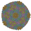
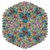
 Map data Map data | Surface rendered view of the asymmetric P22 Virion | |||||||||
 Sample Sample |
| |||||||||
| Biological species |  Enterobacteria phage P22 (virus) Enterobacteria phage P22 (virus) | |||||||||
| Method | single particle reconstruction / cryo EM / Resolution: 17.0 Å | |||||||||
 Authors Authors | Lander GC / Tang L / Casjens SR / Gilcrease EB / Prevelige P / Poliakov A / Potter CS / Carragher B / Johnson JE | |||||||||
 Citation Citation | Journal: Science / Year: 2006 Title: The structure of an infectious P22 virion shows the signal for headful DNA packaging. Authors: Gabriel C Lander / Liang Tang / Sherwood R Casjens / Eddie B Gilcrease / Peter Prevelige / Anton Poliakov / Clinton S Potter / Bridget Carragher / John E Johnson /  Abstract: Bacteriophages, herpesviruses, and other large double-stranded DNA (dsDNA) viruses contain molecular machines that pump DNA into preassembled procapsids, generating internal capsid pressures ...Bacteriophages, herpesviruses, and other large double-stranded DNA (dsDNA) viruses contain molecular machines that pump DNA into preassembled procapsids, generating internal capsid pressures exceeding, by 10-fold, that of bottled champagne. A 17 angstrom resolution asymmetric reconstruction of the infectious P22 virion reveals that tightly spooled DNA about the portal dodecamer forces a conformation that is significantly different from that observed in isolated portals assembled from ectopically expressed protein. We propose that the tight dsDNA spooling activates the switch that signals the headful chromosome packing density to the particle exterior. | |||||||||
| History |
|
- Structure visualization
| Movie |
 Movie viewer Movie viewer |
|---|---|
| Structure viewer | EM map: SurfViewMolmilJmol/JSmol |
| Supplemental images |

- Downloads & links
-EMDB archive
| Map data | emd_1220.map.gz | 59.1 MB | EMDB map data format | |
|---|---|---|---|---|
| Header (meta data) | emd-1220-v30.xmlemd-1220.xml | 9.5 KB 9.5 KB | Display Display | EMDB header |
| Images | 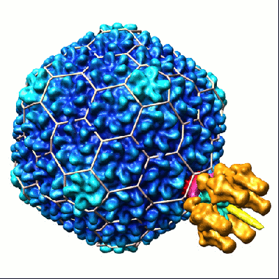 1220.gif 1220.gif | 79.7 KB | ||
| Archive directory |  http://ftp.pdbj.org/pub/emdb/structures/EMD-1220ftp://ftp.pdbj.org/pub/emdb/structures/EMD-1220 http://ftp.pdbj.org/pub/emdb/structures/EMD-1220ftp://ftp.pdbj.org/pub/emdb/structures/EMD-1220 | HTTPS FTP |
-Validation report
| Summary document | emd_1220_validation.pdf.gz | 255.4 KB | Display | EMDB validaton report |
|---|---|---|---|---|
| Full document | emd_1220_full_validation.pdf.gz | 254.5 KB | Display | |
| Data in XML | emd_1220_validation.xml.gz | 6.6 KB | Display | |
| Arichive directory | https://ftp.pdbj.org/pub/emdb/validation_reports/EMD-1220ftp://ftp.pdbj.org/pub/emdb/validation_reports/EMD-1220 | HTTPS FTP |
-Related structure data
| Similar structure data |
|---|

-Links
| EMDB pages | EMDB (EBI/PDBe) / EMDataResource |
|---|
-Map
| File | Download / File: emd_1220.map.gz / Format: CCP4 / Size: 62.5 MB / Type: IMAGE STORED AS FLOATING POINT NUMBER (4 BYTES) | ||||||||||||||||||||||||||||||||||||||||||||||||||||||||||||||||||||
|---|---|---|---|---|---|---|---|---|---|---|---|---|---|---|---|---|---|---|---|---|---|---|---|---|---|---|---|---|---|---|---|---|---|---|---|---|---|---|---|---|---|---|---|---|---|---|---|---|---|---|---|---|---|---|---|---|---|---|---|---|---|---|---|---|---|---|---|---|---|
| Annotation | Surface rendered view of the asymmetric P22 Virion | ||||||||||||||||||||||||||||||||||||||||||||||||||||||||||||||||||||
| Voxel size | X=Y=Z: 4.04 Å | ||||||||||||||||||||||||||||||||||||||||||||||||||||||||||||||||||||
| Density |
| ||||||||||||||||||||||||||||||||||||||||||||||||||||||||||||||||||||
| Symmetry | Space group: 1 | ||||||||||||||||||||||||||||||||||||||||||||||||||||||||||||||||||||
| Details | EMDB XML:
CCP4 map header:
| ||||||||||||||||||||||||||||||||||||||||||||||||||||||||||||||||||||
-Supplemental data
- Sample components
-Entire : P22 Bacteriophage Virion
| Entire | Name: P22 Bacteriophage Virion |
|---|---|
| Components |
|
-Supramolecule #1000: P22 Bacteriophage Virion
| Supramolecule | Name: P22 Bacteriophage Virion / type: sample / ID: 1000 / Number unique components: 1 |
|---|
-Supramolecule #1: Enterobacteria phage P22
| Supramolecule | Name: Enterobacteria phage P22 / type: virus / ID: 1 / NCBI-ID: 10754 / Sci species name: Enterobacteria phage P22 / Virus type: VIRION / Virus isolate: STRAIN / Virus enveloped: No / Virus empty: No |
|---|---|
| Host (natural) | Organism:  Salmonella enterica (bacteria) / synonym: BACTERIA(EUBACTERIA) Salmonella enterica (bacteria) / synonym: BACTERIA(EUBACTERIA) |
| Virus shell | Shell ID: 1 / Name: gp5 / Diameter: 600 Å / T number (triangulation number): 7 |
-Experimental details
-Structure determination
| Method | cryo EM |
|---|---|
 Processing Processing | single particle reconstruction |
| Aggregation state | particle |
-Sample preparation
| Concentration | 10 mg/mL |
|---|---|
| Buffer | pH: 7.4 / Details: 10mM Tris-HCL, 1mM MgCl2 |
| Grid | Details: Quantifoil 2/2 400 mesh grid |
| Vitrification | Cryogen name: ETHANE / Chamber humidity: 100 % / Chamber temperature: 4.2 K / Instrument: OTHER / Details: Vitrification instrument: FEI Vitrobot / Method: Blot for 4 seconds before plunging |
- Electron microscopy
| Microscope | FEI TECNAI F20 |
|---|---|
| Temperature | Min: 4.5 K / Max: 4.8 K / Average: 4.5 K |
| Alignment procedure | Legacy - Astigmatism: Objective lens astigmatism was corrected automatically at 50,000 times magnification |
| Specialist optics | Energy filter - Name: FEI / Energy filter - Lower energy threshold: 0.0 eV / Energy filter - Upper energy threshold: 25.0 eV |
| Details | images collected automatically with LEGINON |
| Image recording | Category: CCD / Film or detector model: GENERIC TVIPS / Number real images: 4899 / Average electron dose: 20 e/Å2 |
| Electron beam | Acceleration voltage: 200 kV / Electron source:  FIELD EMISSION GUN FIELD EMISSION GUN |
| Electron optics | Calibrated magnification: 80000 / Illumination mode: FLOOD BEAM / Imaging mode: BRIGHT FIELD / Cs: 2 mm / Nominal defocus max: 4.5 µm / Nominal defocus min: 0.5 µm / Nominal magnification: 80000 |
| Sample stage | Specimen holder: Eucentric / Specimen holder model: GATAN LIQUID NITROGEN |
| Experimental equipment |  Model: Tecnai F20 / Image courtesy: FEI Company |
-Image processing
| Details | Particles selected manually, CTF estimation was performed using Automated CTF-Estimation Matlab package (ACE) |
|---|---|
| CTF correction | Details: Each micrograph |
| Final reconstruction | Applied symmetry - Point group: C1 (asymmetric) / Algorithm: OTHER / Resolution.type: BY AUTHOR / Resolution: 17.0 Å / Resolution method: FSC 0.5 CUT-OFF / Software - Name: SPIDER, EMAN / Number images used: 22968 |
