 Movie
Movie Controller

[English] 日本語
 Yorodumi
- EMDB-1166: Cryo-EM reconstruction of dengue virus in complex with the carboh... -
+ Open data

- Basic information
| Entry | Database: EMDB / ID: EMD-1166 | |||||||||
|---|---|---|---|---|---|---|---|---|---|---|
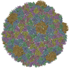
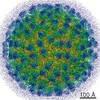
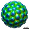
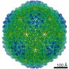
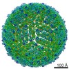
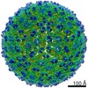
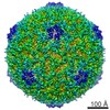
| Title | Cryo-EM reconstruction of dengue virus in complex with the carbohydrate recognition domain of DC-SIGN. | |||||||||
 Map data Map data | EM map of dengue virus in complex with CRD domain of DC-SIGN | |||||||||
 Sample Sample |
| |||||||||
| Function / homology |  Function and homology information Function and homology informationB cell adhesion /  cell-cell recognition / peptide antigen transport / modulation by virus of host process / intracellular transport of virus / Butyrophilin (BTN) family interactions / positive regulation of viral life cycle / host cell / virion binding / heterophilic cell-cell adhesion via plasma membrane cell adhesion molecules ...B cell adhesion / cell-cell recognition / peptide antigen transport / modulation by virus of host process / intracellular transport of virus / Butyrophilin (BTN) family interactions / positive regulation of viral life cycle / host cell / virion binding / heterophilic cell-cell adhesion via plasma membrane cell adhesion molecules / leukocyte cell-cell adhesion / regulation of T cell proliferation / symbiont-mediated suppression of host JAK-STAT cascade via inhibition of host TYK2 activity / host cell mitochondrion / stimulatory C-type lectin receptor signaling pathway / mannose binding / antigen processing and presentation / symbiont-mediated suppression of host JAK-STAT cascade via inhibition of STAT2 activity / positive regulation of T cell proliferation / symbiont-mediated suppression of host cytoplasmic pattern recognition receptor signaling pathway via inhibition of MAVS activity / CD209 (DC-SIGN) signaling / ribonucleoside triphosphate phosphatase activity / viral genome replication / endocytosis / : / peptide antigen binding / viral capsid / double-stranded RNA binding / protein complex oligomerization / virus receptor activity / monoatomic ion channel activity / carbohydrate binding / clathrin-dependent endocytosis of virus by host cell / mRNA (nucleoside-2'-O-)-methyltransferase activity / mRNA 5'-cap (guanine-N7-)-methyltransferase activity / RNA helicase activity / adaptive immune response / membrane => GO:0016020 / host cell endoplasmic reticulum membrane / protein dimerization activity / intracellular signal transduction / immune response / induction by virus of host autophagy / symbiont entry into host cell / viral RNA genome replication / external side of plasma membrane / RNA-dependent RNA polymerase activity / serine-type endopeptidase activity / innate immune response / fusion of virus membrane with host endosome membrane / viral envelope / symbiont-mediated suppression of host type I interferon-mediated signaling pathway / host cell nucleus / structural molecule activity / virion attachment to host cell / virion membrane / cell surface / proteolysis / extracellular region / ATP binding / membrane / metal ion binding / plasma membrane / cytoplasm cell-cell recognition / peptide antigen transport / modulation by virus of host process / intracellular transport of virus / Butyrophilin (BTN) family interactions / positive regulation of viral life cycle / host cell / virion binding / heterophilic cell-cell adhesion via plasma membrane cell adhesion molecules ...B cell adhesion / cell-cell recognition / peptide antigen transport / modulation by virus of host process / intracellular transport of virus / Butyrophilin (BTN) family interactions / positive regulation of viral life cycle / host cell / virion binding / heterophilic cell-cell adhesion via plasma membrane cell adhesion molecules / leukocyte cell-cell adhesion / regulation of T cell proliferation / symbiont-mediated suppression of host JAK-STAT cascade via inhibition of host TYK2 activity / host cell mitochondrion / stimulatory C-type lectin receptor signaling pathway / mannose binding / antigen processing and presentation / symbiont-mediated suppression of host JAK-STAT cascade via inhibition of STAT2 activity / positive regulation of T cell proliferation / symbiont-mediated suppression of host cytoplasmic pattern recognition receptor signaling pathway via inhibition of MAVS activity / CD209 (DC-SIGN) signaling / ribonucleoside triphosphate phosphatase activity / viral genome replication / endocytosis / : / peptide antigen binding / viral capsid / double-stranded RNA binding / protein complex oligomerization / virus receptor activity / monoatomic ion channel activity / carbohydrate binding / clathrin-dependent endocytosis of virus by host cell / mRNA (nucleoside-2'-O-)-methyltransferase activity / mRNA 5'-cap (guanine-N7-)-methyltransferase activity / RNA helicase activity / adaptive immune response / membrane => GO:0016020 / host cell endoplasmic reticulum membrane / protein dimerization activity / intracellular signal transduction / immune response / induction by virus of host autophagy / symbiont entry into host cell / viral RNA genome replication / external side of plasma membrane / RNA-dependent RNA polymerase activity / serine-type endopeptidase activity / innate immune response / fusion of virus membrane with host endosome membrane / viral envelope / symbiont-mediated suppression of host type I interferon-mediated signaling pathway / host cell nucleus / structural molecule activity / virion attachment to host cell / virion membrane / cell surface / proteolysis / extracellular region / ATP binding / membrane / metal ion binding / plasma membrane / cytoplasmSimilarity search - Function | |||||||||
| Biological species |  Homo sapiens (human) / Dengue-2 Homo sapiens (human) / Dengue-2 | |||||||||
| Method | single particle reconstruction / cryo EM / Resolution: 25.0 Å | |||||||||
 Authors Authors | Pokidysheva E / Zhang Y / Battisti AJ / Bator-Kelly CM / Chipman PR / Xiao C / Gregorio GG / Hendrickson WA / Kuhn RJ / Rossmann MG | |||||||||
 Citation Citation | Journal: Cell / Year: 2006 Title: Cryo-EM reconstruction of dengue virus in complex with the carbohydrate recognition domain of DC-SIGN. Authors: Elena Pokidysheva / Ying Zhang / Anthony J Battisti / Carol M Bator-Kelly / Paul R Chipman / Chuan Xiao / G Glenn Gregorio / Wayne A Hendrickson / Richard J Kuhn / Michael G Rossmann /  Abstract: Dengue virus (DENV) is a significant human pathogen that causes millions of infections and results in about 24,000 deaths each year. Dendritic cell-specific ICAM3 grabbing nonintegrin (DC-SIGN), ...Dengue virus (DENV) is a significant human pathogen that causes millions of infections and results in about 24,000 deaths each year. Dendritic cell-specific ICAM3 grabbing nonintegrin (DC-SIGN), abundant in immature dendritic cells, was previously reported as being an ancillary receptor interacting with the surface of DENV. The structure of DENV in complex with the carbohydrate recognition domain (CRD) of DC-SIGN was determined by cryo-electron microscopy at 25 A resolution. One CRD monomer was found to bind to two glycosylation sites at Asn67 of two neighboring glycoproteins in each icosahedral asymmetric unit, leaving the third Asn67 residue vacant. The vacancy at the third Asn67 site is a result of the nonequivalence of the glycoprotein environments, leaving space for the primary receptor binding to domain III of E. The use of carbohydrate moieties for receptor binding sites suggests a mechanism for avoiding immune surveillance. | |||||||||
| History |
|
- Structure visualization
| Movie |
Movie viewer |
|---|---|
| Structure viewer | EM map: SurfViewMolmilJmol/JSmol |
| Supplemental images |

- Downloads & links
-EMDB archive
| Map data | emd_1166.map.gz | 4.9 MB | EMDB map data format | |
|---|---|---|---|---|
| Header (meta data) | emd-1166-v30.xmlemd-1166.xml | 11.5 KB 11.5 KB | Display Display | EMDB header |
| Images |  1166.gif 1166.gif | 12 KB | ||
| Archive directory |  http://ftp.pdbj.org/pub/emdb/structures/EMD-1166ftp://ftp.pdbj.org/pub/emdb/structures/EMD-1166 http://ftp.pdbj.org/pub/emdb/structures/EMD-1166ftp://ftp.pdbj.org/pub/emdb/structures/EMD-1166 | HTTPS FTP |
-Related structure data
| Related structure data |  2b6bMC  1167C M: atomic model generated by this map C: citing same article ( |
|---|---|
| Similar structure data |

-Links
| EMDB pages | EMDB (EBI/PDBe) / EMDataResource |
|---|---|
| Related items in Molecule of the Month |


-Map
| File | Download / File: emd_1166.map.gz / Format: CCP4 / Size: 13.6 MB / Type: IMAGE STORED AS FLOATING POINT NUMBER (4 BYTES) | ||||||||||||||||||||||||||||||||||||||||||||||||||||||||||||||||||||
|---|---|---|---|---|---|---|---|---|---|---|---|---|---|---|---|---|---|---|---|---|---|---|---|---|---|---|---|---|---|---|---|---|---|---|---|---|---|---|---|---|---|---|---|---|---|---|---|---|---|---|---|---|---|---|---|---|---|---|---|---|---|---|---|---|---|---|---|---|---|
| Annotation | EM map of dengue virus in complex with CRD domain of DC-SIGN | ||||||||||||||||||||||||||||||||||||||||||||||||||||||||||||||||||||
| Voxel size | X=Y=Z: 4.34 Å | ||||||||||||||||||||||||||||||||||||||||||||||||||||||||||||||||||||
| Density |
| ||||||||||||||||||||||||||||||||||||||||||||||||||||||||||||||||||||
| Symmetry | Space group: 1 | ||||||||||||||||||||||||||||||||||||||||||||||||||||||||||||||||||||
| Details | EMDB XML:
CCP4 map header:
| ||||||||||||||||||||||||||||||||||||||||||||||||||||||||||||||||||||
-Supplemental data
- Sample components
-Entire : Dengue virus complexed with CRD domain of DC-SIGN
| Entire | Name: Dengue virus complexed with CRD domain of DC-SIGN |
|---|---|
| Components |
|
-Supramolecule #1000: Dengue virus complexed with CRD domain of DC-SIGN
| Supramolecule | Name: Dengue virus complexed with CRD domain of DC-SIGN / type: sample / ID: 1000 / Details: Calcium should be present in the sample. / Oligomeric state: one monomer of CRD binds virus ico / Number unique components: 2 |
|---|---|
| Molecular weight | Theoretical: 12 MDa |
-Supramolecule #1: Dengue-2
| Supramolecule | Name: Dengue-2 / type: virus / ID: 1 / Sci species name: Dengue-2 / Virus type: VIRION / Virus isolate: STRAIN / Virus enveloped: Yes / Virus empty: No |
|---|---|
| Host (natural) | Organism: Homo sapiens (human) / synonym: VERTEBRATES |
| Molecular weight | Experimental: 11 MDa / Theoretical: 11 MDa |
| Virus shell | Shell ID: 1 / Name: outer shell, T is not applicable / Diameter: 500 Å / T number (triangulation number): 3 |
-Macromolecule #1: CRD domain of DC-SIGN
| Macromolecule | Name: CRD domain of DC-SIGN / type: protein_or_peptide / ID: 1 Details: expressed insoluble in the inclusion bodies. Refolded. Oligomeric state: monomer / Recombinant expression: Yes |
|---|---|
| Source (natural) | Organism: Homo sapiens (human) / synonym: Human / Tissue: dendritic cells / Cell: bacteria BL21-DE3 / Location in cell: cell membrane, extracellular matrix |
| Molecular weight | Experimental: 18 KDa / Theoretical: 18 KDa |
| Recombinant expression | Organism:  Escherichia coli (E. coli) / Recombinant plasmid: pet 28 Escherichia coli (E. coli) / Recombinant plasmid: pet 28 |
-Experimental details
-Structure determination
| Method | cryo EM |
|---|---|
 Processing Processing | single particle reconstruction |
| Aggregation state | particle |
-Sample preparation
| Concentration | 2 mg/mL |
|---|---|
| Buffer | pH: 7.5 Details: 50mM Tris 50mM NaCl 0.5 mM EDTA, 5 mM CaCl2, pH 7.5 |
| Grid | Details: 400 mesh copper grid |
| Vitrification | Cryogen name: ETHANE / Instrument: HOMEMADE PLUNGER Details: Vitrification instrument: Guillotine-style plunge freezeing device Method: Small aliquots of sample were applied to 400 mesh copper grids coated with holey carbon film and rapidly frozen by plunging into an ethane slush |
- Electron microscopy
| Microscope | FEI/PHILIPS CM300FEG/T |
|---|---|
| Electron beam | Acceleration voltage: 300 kV / Electron source: FIELD EMISSION GUN |
| Electron optics | Calibrated magnification: 33000 / Illumination mode: OTHER / Imaging mode: BRIGHT FIELDBright-field microscopy / Cs: 2.0 mm / Nominal defocus max: 3.0 µm / Nominal defocus min: 1.0 µm / Nominal magnification: 33000 |
| Sample stage | Specimen holder: Cryo / Specimen holder model: GATAN LIQUID NITROGEN |
| Temperature | Average: 103 K |
| Alignment procedure | Legacy - Astigmatism: objective lens astigmatism was corrected at 98,000 times magnification |
| Date | Nov 15, 2004 |
| Image recording | Category: FILM / Film or detector model: KODAK SO-163 FILM / Digitization - Scanner: ZEISS SCAI / Digitization - Sampling interval: 7 µm / Number real images: 45 / Average electron dose: 11.8 e/Å2 / Od range: 1.1 / Bits/pixel: 8 |
-Image processing
| CTF correction | Details: each particle |
|---|---|
| Final angle assignment | Details: SPIDER theta 36.4 degrees, phi 72 degrees |
| Final reconstruction | Applied symmetry - Point group: I (icosahedral) / Algorithm: OTHER / Resolution.type: BY AUTHOR / Resolution: 25.0 Å / Resolution method: FSC 0.5 CUT-OFF / Software - Name: spider / Number images used: 830 |
| Details | the particles were selected manualy |
