 Movie
Movie Controller

[English] 日本語
 Yorodumi
- EMDB-1036: Structure of a fast kinesin: implications for ATPase mechanism an... -
+ Open data

- Basic information
| Entry | Database: EMDB / ID: EMD-1036 | |||||||||
|---|---|---|---|---|---|---|---|---|---|---|
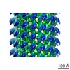
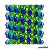
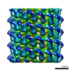
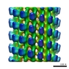
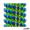
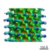
| Title | Structure of a fast kinesin: implications for ATPase mechanism and interactions with microtubules. | |||||||||
 Map data Map data | Neurospora crassa kinesin monomer (nK355) AMP-PNP state | |||||||||
 Sample Sample |
| |||||||||
| Biological species |   Neurospora crassa (fungus) Neurospora crassa (fungus) | |||||||||
| Method | helical reconstruction / cryo EM / negative staining / Resolution: 25.0 Å | |||||||||
 Authors Authors | Krebs A | |||||||||
 Citation Citation | Journal: EMBO J / Year: 2001 Title: Structure of a fast kinesin: implications for ATPase mechanism and interactions with microtubules. Authors: Y H Song / A Marx / J Müller / G Woehlke / M Schliwa / A Krebs / A Hoenger / E Mandelkow /  Abstract: We determined the crystal structure of the motor domain of the fast fungal kinesin from Neurospora crassa (NcKin). The structure has several unique features. (i) Loop 11 in the switch 2 region is ...We determined the crystal structure of the motor domain of the fast fungal kinesin from Neurospora crassa (NcKin). The structure has several unique features. (i) Loop 11 in the switch 2 region is ordered and enables one to describe the complete nucleotide-binding pocket, including three inter-switch salt bridges between switch 1 and 2. (ii) Loop 9 in the switch 1 region bends outwards, making the nucleotide-binding pocket very wide. The displacement in switch 1 resembles that of the G-protein ras complexed with its guanosine nucleotide exchange factor. (iii) Loop 5 in the entrance to the nucleotide-binding pocket is remarkably long and interacts with the ribose of ATP. (iv) The linker and neck region is not well defined, indicating that it is mobile. (v) Image reconstructions of ice-embedded microtubules decorated with NcKin show that it interacts with several tubulin subunits, including a central beta-tubulin monomer and the two flanking alpha-tubulin monomers within the microtubule protofilament. Comparison of NcKin with other kinesins, myosin and G-proteins suggests that the rate-limiting step of ADP release is accelerated in the fungal kinesin and accounts for the unusually high velocity and ATPase activity. | |||||||||
| History |
|
- Structure visualization
| Movie |
 Movie viewer Movie viewer |
|---|---|
| Structure viewer | EM map: SurfViewMolmilJmol/JSmol |
| Supplemental images |
- Downloads & links
-EMDB archive
| Map data | emd_1036.map.gz | 2.8 MB | EMDB map data format | |
|---|---|---|---|---|
| Header (meta data) | emd-1036-v30.xmlemd-1036.xml | 9.7 KB 9.7 KB | Display Display | EMDB header |
| Images |  1036.gif 1036.gif | 64.9 KB | ||
| Archive directory |  http://ftp.pdbj.org/pub/emdb/structures/EMD-1036ftp://ftp.pdbj.org/pub/emdb/structures/EMD-1036 http://ftp.pdbj.org/pub/emdb/structures/EMD-1036ftp://ftp.pdbj.org/pub/emdb/structures/EMD-1036 | HTTPS FTP |
-Related structure data


-Links
| EMDB pages | EMDB (EBI/PDBe) / EMDataResource |
|---|
-Map
| File | Download / File: emd_1036.map.gz / Format: CCP4 / Size: 3.7 MB / Type: IMAGE STORED AS FLOATING POINT NUMBER (4 BYTES) | ||||||||||||||||||||||||||||||||||||||||||||||||||||||||||||
|---|---|---|---|---|---|---|---|---|---|---|---|---|---|---|---|---|---|---|---|---|---|---|---|---|---|---|---|---|---|---|---|---|---|---|---|---|---|---|---|---|---|---|---|---|---|---|---|---|---|---|---|---|---|---|---|---|---|---|---|---|---|
| Annotation | Neurospora crassa kinesin monomer (nK355) AMP-PNP state | ||||||||||||||||||||||||||||||||||||||||||||||||||||||||||||
| Voxel size | X=Y=Z: 5.526 Å | ||||||||||||||||||||||||||||||||||||||||||||||||||||||||||||
| Density |
| ||||||||||||||||||||||||||||||||||||||||||||||||||||||||||||
| Symmetry | Space group: 1 | ||||||||||||||||||||||||||||||||||||||||||||||||||||||||||||
| Details | EMDB XML:
CCP4 map header:
| ||||||||||||||||||||||||||||||||||||||||||||||||||||||||||||
-Supplemental data
- Sample components
-Entire : neurospora crassa kinesin monomer
| Entire | Name: neurospora crassa kinesin monomer |
|---|---|
| Components |
|
-Supramolecule #1000: neurospora crassa kinesin monomer
| Supramolecule | Name: neurospora crassa kinesin monomer / type: sample / ID: 1000 / Oligomeric state: monomer / Number unique components: 2 |
|---|
-Macromolecule #1: neurospora crassakinesin
| Macromolecule | Name: neurospora crassakinesin / type: protein_or_peptide / ID: 1 / Name.synonym: molecular motor / Number of copies: 1 / Oligomeric state: monomer / Recombinant expression: Yes / Database: NCBI |
|---|---|
| Source (natural) | Organism: Neurospora crassa (fungus) / synonym: neurospora |
-Macromolecule #2: tubulin
| Macromolecule | Name: tubulin / type: protein_or_peptide / ID: 2 / Name.synonym: microtubules / Number of copies: 1 / Oligomeric state: hetero dimer / Recombinant expression: No / Database: NCBI |
|---|---|
| Source (natural) | Organism: Neurospora crassa (fungus) / synonym: neurospora / Tissue: brain / Cell: neuronal cells / Location in cell: cytoplasm |
-Experimental details
-Structure determination
| Method | negative staining, cryo EM |
|---|---|
 Processing Processing | helical reconstruction |
| Aggregation state | filament |
-Sample preparation
| Concentration | 0.5 mg/mL |
|---|---|
| Buffer | pH: 6.9 Details: Pipes 20mM, 50 mM NaCl, 5mM mgcl,1mM Mg-ATP, 20um taxol. |
| Staining | Type: NEGATIVE / Details: ice-embedded |
| Grid | Details: holey grids |
| Vitrification | Cryogen name: PROPANE / Chamber temperature: 93 K / Instrument: HOMEMADE PLUNGER / Details: Vitrification instrument: self made |
- Electron microscopy
| Microscope | FEI/PHILIPS CM120T |
|---|---|
| Electron beam | Acceleration voltage: 100 kV / Electron source: LAB6 |
| Electron optics | Illumination mode: FLOOD BEAM / Imaging mode: BRIGHT FIELDBright-field microscopy / Nominal defocus max: 2.0 µm / Nominal defocus min: 1.5 µm / Nominal magnification: 37000 |
| Sample stage | Specimen holder: side entry / Specimen holder model: GATAN LIQUID NITROGEN |
| Image recording | Category: FILM / Film or detector model: KODAK SO-163 FILM / Digitization - Scanner: ZEISS SCAI / Digitization - Sampling interval: 21 µm / Number real images: 18 / Average electron dose: 5 e/Å2 / Bits/pixel: 16 |
| Tilt angle min | 0 |
| Tilt angle max | 0 |
-Image processing
| Final reconstruction | Applied symmetry - Helical parameters - Axial symmetry: C1 (asymmetric) Algorithm: OTHER / Resolution.type: BY AUTHOR / Resolution: 25.0 Å / Resolution method: FSC 0.5 CUT-OFF / Software - Name: Phoelix, Suprim Details: Final maps from 36 averaged datasets = 18 helical tubes |
|---|
