 Movie
Movie Controller

[English] 日本語
 Yorodumi
- PDB-5grh: Crystal structure of the alpha gamma heterodimer of human IDH3 in... -
+ Open data

- Basic information
| Entry | Database: PDB / ID: 5grh | ||||||||||||
|---|---|---|---|---|---|---|---|---|---|---|---|---|---|
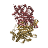
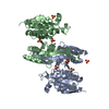
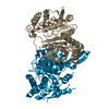
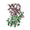
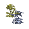
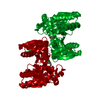
| Title | Crystal structure of the alpha gamma heterodimer of human IDH3 in complex with Mg(2+) | ||||||||||||
 Components Components |
| ||||||||||||
 Keywords Keywords |  OXIDOREDUCTASE / apo-form / isocitrate dehydrogenase OXIDOREDUCTASE / apo-form / isocitrate dehydrogenase | ||||||||||||
| Function / homology |  Function and homology information: / isocitrate dehydrogenase (NAD+) / isocitrate dehydrogenase (NAD+) activity / Citric acid cycle (TCA cycle) / isocitrate metabolic process / Mitochondrial protein import / tricarboxylic acid cycle / NAD binding / carbohydrate metabolic process / mitochondrial matrix ...: / isocitrate dehydrogenase (NAD+) / isocitrate dehydrogenase (NAD+) activity / Citric acid cycle (TCA cycle) / isocitrate metabolic process / Mitochondrial protein import / tricarboxylic acid cycle / NAD binding / carbohydrate metabolic process / mitochondrial matrix / nucleolus / magnesium ion binding / mitochondrion / ATP binding / nucleus Function and homology information: / isocitrate dehydrogenase (NAD+) / isocitrate dehydrogenase (NAD+) activity / Citric acid cycle (TCA cycle) / isocitrate metabolic process / Mitochondrial protein import / tricarboxylic acid cycle / NAD binding / carbohydrate metabolic process / mitochondrial matrix ...: / isocitrate dehydrogenase (NAD+) / isocitrate dehydrogenase (NAD+) activity / Citric acid cycle (TCA cycle) / isocitrate metabolic process / Mitochondrial protein import / tricarboxylic acid cycle / NAD binding / carbohydrate metabolic process / mitochondrial matrix / nucleolus / magnesium ion binding / mitochondrion / ATP binding / nucleusSimilarity search - Function | ||||||||||||
| Biological species |  Homo sapiens (human) Homo sapiens (human) | ||||||||||||
| Method | X-RAY DIFFRACTION / SYNCHROTRON / MOLECULAR REPLACEMENT / Resolution: 2.8 Å | ||||||||||||
 Authors Authors | Ma, T. / Peng, Y. | ||||||||||||
| Funding support |  China, 3items China, 3items
| ||||||||||||
 Citation Citation | Journal: Sci Rep / Year: 2017 Title: Molecular mechanism of the allosteric regulation of the alpha gamma heterodimer of human NAD-dependent isocitrate dehydrogenase. Authors: Ma, T. / Peng, Y. / Huang, W. / Ding, J. | ||||||||||||
| History |
|
- Structure visualization
| Structure viewer | Molecule: MolmilJmol/JSmol |
|---|
- Downloads & links
-Download
| PDBx/mmCIF format | 5grh.cif.gz | 139.3 KB | Display | PDBx/mmCIF format |
|---|---|---|---|---|
| PDB format | pdb5grh.ent.gz | 107.5 KB | Display | PDB format |
| PDBx/mmJSON format | 5grh.json.gz | Tree view | PDBx/mmJSON format | |
| Others |  Other downloads Other downloads |
-Validation report
| Arichive directory | https://data.pdbj.org/pub/pdb/validation_reports/gr/5grhftp://data.pdbj.org/pub/pdb/validation_reports/gr/5grh | HTTPS FTP |
|---|
-Related structure data
| Related structure data |  5greC  5grfC  5griC 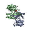 5grlC 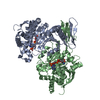 1t09S C: citing same article ( S: Starting model for refinement |
|---|---|
| Similar structure data |


-Links
 PDBj
PDBj

- Assembly
| Deposited unit | 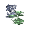
| ||||||||
|---|---|---|---|---|---|---|---|---|---|
| 1 |
| ||||||||
| Unit cell |
|
-Components
| #1: Protein | Mass: 36682.176 Da / Num. of mol.: 1 Source method: isolated from a genetically manipulated source Source: (gene. exp.) Homo sapiens (human) / Gene: IDH3A / Production host:  Escherichia coli BL21(DE3) (bacteria) / Strain (production host): BL21(DE3) Escherichia coli BL21(DE3) (bacteria) / Strain (production host): BL21(DE3)References: UniProt: P50213, isocitrate dehydrogenase (NAD+) |
|---|---|
| #2: Protein | Mass: 38867.523 Da / Num. of mol.: 1 Source method: isolated from a genetically manipulated source Source: (gene. exp.) Homo sapiens (human) / Gene: IDH3G / Production host: Escherichia coli BL21(DE3) (bacteria) / Strain (production host): BL21(DE3)References: UniProt: P51553, isocitrate dehydrogenase (NAD+) |
| #3: Chemical | ChemComp-MG /   Mass: 24.305 Da / Num. of mol.: 1 / Source method: obtained synthetically / Formula: Mg Mass: 24.305 Da / Num. of mol.: 1 / Source method: obtained synthetically / Formula: Mg |
-Experimental details
-Experiment
| Experiment | Method: X-RAY DIFFRACTION / Number of used crystals: 1 |
|---|
- Sample preparation
| Crystal | Density Matthews: 3.83 Å3/Da / Density % sol: 67.92 % |
|---|---|
| Crystal grow | Temperature: 293 K / Method: vapor diffusion, hanging drop Details: 0.1 M HEPES-Na, pH7.5, 50 mM MgCl2, 30%(v/v) PEGMME 550 |
-Data collection
| Diffraction | Mean temperature: 100 K |
|---|---|
| Diffraction source | Source: SYNCHROTRON / Site: SSRF / Beamline: BL19U1 / Wavelength: 1 Å |
| Detector | Type: DECTRIS PILATUS3 6M / Detector: PIXEL / Date: Jun 2, 2015 |
| Radiation | Protocol: SINGLE WAVELENGTH / Monochromatic (M) / Laue (L): M / Scattering type: x-ray |
| Radiation wavelength | Wavelength: 1 Å / Relative weight: 1 |
| Reflection | Resolution: 2.8→19.71 Å / Num. obs: 28718 / % possible obs: 98.9 % / Redundancy: 10.8 % / Net I/σ(I): 9.6 |
| Reflection shell | Resolution: 2.8→2.9 Å |
- Processing
| Software |
| ||||||||||||||||||||||||||||||||||||||||||||||||||||||||||||
|---|---|---|---|---|---|---|---|---|---|---|---|---|---|---|---|---|---|---|---|---|---|---|---|---|---|---|---|---|---|---|---|---|---|---|---|---|---|---|---|---|---|---|---|---|---|---|---|---|---|---|---|---|---|---|---|---|---|---|---|---|---|
| Refinement | Method to determine structure: MOLECULAR REPLACEMENT Starting model: 1T09 Resolution: 2.8→19.71 Å / Cor.coef. Fo:Fc: 0.928 / Cor.coef. Fo:Fc free: 0.9 / SU B: 12.47 / SU ML: 0.234 / Cross valid method: THROUGHOUT / σ(F): 0 / ESU R: 0.116 / ESU R Free: 0.068 Details: HYDROGENS HAVE BEEN ADDED IN THE RIDING POSITIONS U VALUES
| ||||||||||||||||||||||||||||||||||||||||||||||||||||||||||||
| Solvent computation | Ion probe radii: 0.8 Å / Shrinkage radii: 0.8 Å / VDW probe radii: 1.2 Å | ||||||||||||||||||||||||||||||||||||||||||||||||||||||||||||
| Displacement parameters | Biso max: 170.32 Å2 / Biso mean: 58.397 Å2 / Biso min: 23.81 Å2
| ||||||||||||||||||||||||||||||||||||||||||||||||||||||||||||
| Refinement step | Cycle: final / Resolution: 2.8→19.71 Å
| ||||||||||||||||||||||||||||||||||||||||||||||||||||||||||||
| Refine LS restraints |
| ||||||||||||||||||||||||||||||||||||||||||||||||||||||||||||
| LS refinement shell | Resolution: 2.8→2.873 Å / Total num. of bins used: 20
|
