- PDB-4g83: Crystal Structure of p73 DNA-Binding Domain Tetramer bound to a F... -
+
Open data
ID or keywords:
Loading...
-
Basic information
Entry
Database: PDB / ID: 4g83
Title
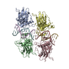
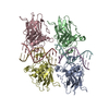
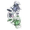
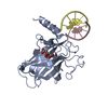
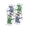
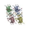
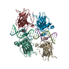
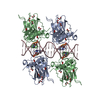
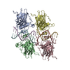
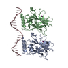
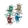
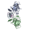
Crystal Structure of p73 DNA-Binding Domain Tetramer bound to a Full Response-Element
Components
Tumor protein p73P73
dna
Keywords
DNA BINDING PROTEIN/DNA / BETA-IMMUNOGLOBULIN FOLD / TUMOR SUPPRESSOR / DNA BINDING PROTEIN-DNA complex
Function / homology
Function and homology information
positive regulation of lung ciliated cell differentiation / negative regulation of cardiac muscle cell proliferation / TP53 Regulates Transcription of Death Receptors and Ligands / Activation of PUMA and translocation to mitochondria / Regulation of TP53 Activity through Association with Co-factors / positive regulation of oligodendrocyte differentiation / TP53 regulates transcription of several additional cell death genes whose specific roles in p53-dependent apoptosis remain uncertain / TP53 Regulates Transcription of Caspase Activators and Caspases / regulation of mitotic cell cycle / TP53 Regulates Transcription of Genes Involved in Cytochrome C Release ...positive regulation of lung ciliated cell differentiation / negative regulation of cardiac muscle cell proliferation / TP53 Regulates Transcription of Death Receptors and Ligands / Activation of PUMA and translocation to mitochondria / Regulation of TP53 Activity through Association with Co-factors / positive regulation of oligodendrocyte differentiation / TP53 regulates transcription of several additional cell death genes whose specific roles in p53-dependent apoptosis remain uncertain / TP53 Regulates Transcription of Caspase Activators and Caspases / regulation of mitotic cell cycle / TP53 Regulates Transcription of Genes Involved in Cytochrome C Release / negative regulation of neuron differentiation / mismatch repair / intrinsic apoptotic signaling pathway in response to DNA damage by p53 class mediator / MDM2/MDM4 family protein binding / response to organonitrogen compound / transcription corepressor binding / kidney development / protein tetramerization / intrinsic apoptotic signaling pathway in response to DNA damage / p53 binding / cell junction / RUNX1 regulates transcription of genes involved in differentiation of HSCs / regulation of gene expression / DNA-binding transcription activator activity, RNA polymerase II-specific / DNA-binding transcription factor binding / RNA polymerase II-specific DNA-binding transcription factor binding / positive regulation of MAPK cascade / transcription cis-regulatory region binding / regulation of cell cycle / DNA-binding transcription factor activity, RNA polymerase II-specific / response to xenobiotic stimulus / cell cycle / positive regulation of apoptotic process / RNA polymerase II cis-regulatory region sequence-specific DNA binding / DNA-binding transcription factor activity / negative regulation of cell population proliferation / intracellular membrane-bounded organelle / DNA damage response / chromatin / regulation of transcription by RNA polymerase II / protein kinase binding / positive regulation of DNA-templated transcription / Golgi apparatus / positive regulation of transcription by RNA polymerase II / nucleoplasm / identical protein binding / metal ion binding / nucleus / cytosol Similarity search - Function
Resolution: 4→45.61 Å / Cor.coef. Fo:Fc: 0.891 / Cor.coef. Fo:Fc free: 0.887 / SU ML: 0.39 / Isotropic thermal model: TLS refinement / σ(F): 1.35 / Phase error: 28.13 / Stereochemistry target values: ML / Details: HYDROGENS HAVE BEEN USED IF PRESENT IN THE INPUT
Rfactor
Num. reflection
% reflection
Rfree
0.3073
202
4.64 %
Rwork
0.2414
-
-
obs
0.2443
4349
99.82 %
all
-
5072
-
Solvent computation
Shrinkage radii: 0.9 Å / VDW probe radii: 1.11 Å / Solvent model: FLAT BULK SOLVENT MODEL
Displacement parameters
Biso mean: 68.022 Å2
Baniso -1
Baniso -2
Baniso -3
1-
0.48 Å2
0 Å2
0 Å2
2-
-
5.45 Å2
0 Å2
3-
-
-
-5.92 Å2
Refinement step
Cycle: LAST / Resolution: 4→45.61 Å
Protein
Nucleic acid
Ligand
Solvent
Total
Num. atoms
3094
410
2
0
3506
Refine LS restraints
Refine-ID
Type
Dev ideal
Number
X-RAY DIFFRACTION
f_bond_d
0.031
3640
X-RAY DIFFRACTION
f_angle_d
3.062
5032
X-RAY DIFFRACTION
f_dihedral_angle_d
20.914
1402
X-RAY DIFFRACTION
f_chiral_restr
0.1
552
X-RAY DIFFRACTION
f_plane_restr
0.014
594
LS refinement shell
Resolution: 4.0002→45.6125 Å
Rfactor
Num. reflection
% reflection
Rfree
0.3073
202
-
Rwork
0.2414
4147
-
obs
-
-
100 %
Refinement TLS params.
Method: refined / Refine-ID: X-RAY DIFFRACTION
ID
L11 (°2)
L12 (°2)
L13 (°2)
L22 (°2)
L23 (°2)
L33 (°2)
S11 (Å °)
S12 (Å °)
S13 (Å °)
S21 (Å °)
S22 (Å °)
S23 (Å °)
S31 (Å °)
S32 (Å °)
S33 (Å °)
T11 (Å2)
T12 (Å2)
T13 (Å2)
T22 (Å2)
T23 (Å2)
T33 (Å2)
Origin x (Å)
Origin y (Å)
Origin z (Å)
1
2.0375
-0.6919
1.0636
2.6688
-0.7298
1.8812
-0.2864
-0.0078
1.4485
-1.1429
0.4491
-0.3277
-0.1134
0.2876
0.5764
0.3483
0.3152
-0.0555
0.5483
0.5118
0.7995
-22.7571
-32.7521
9.3385
2
5.066
-0.4275
-2.3738
1.6489
0.9162
2.5592
-0.5233
0.4095
-0.5928
-0.5105
-0.2792
-0.1755
1.3492
0.0305
-0.2566
1.3344
0.166
0.0833
0.6077
0.0644
0.5611
-22.7099
-32.7407
8.032
3
2.168
1.1984
-0.4306
8.4996
-3.9227
2.8918
-0.542
0.2087
-0.4675
-0.674
-0.4141
-2.9198
0.1813
0.5368
-0.3165
0.1596
-0.257
-0.1057
0.3177
-0.0777
0.1433
-11.4072
-8.0051
13.2438
4
3.5469
-2.5135
-0.9597
7.3234
1.8799
2.981
-0.1748
-0.4719
-2.0314
1.7329
-0.3354
4.4161
-0.2336
-0.1539
0.0291
-0.0088
-0.0335
-0.4926
0.4532
-0.1318
-1.2789
-49.4701
-28.0807
3.7272
Refinement TLS group
ID
Refine-ID
Refine TLS-ID
Selection details
Auth asym-ID
Auth seq-ID
1
X-RAY DIFFRACTION
1
( CHAINEANDRESID400:409 )
E
400 - 409
2
X-RAY DIFFRACTION
2
( CHAINFANDRESID410:419 )
F
410 - 419
3
X-RAY DIFFRACTION
3
( CHAINAANDRESID1:310 )
A
1 - 310
4
X-RAY DIFFRACTION
4
( CHAINBANDRESID113:310 )
B
113 - 310
+
About Yorodumi
-
News
-
Feb 9, 2022. New format data for meta-information of EMDB entries
New format data for meta-information of EMDB entries
Version 3 of the EMDB header file is now the official format.
The previous official version 1.9 will be removed from the archive.
In the structure databanks used in Yorodumi, some data are registered as the other names, "COVID-19 virus" and "2019-nCoV". Here are the details of the virus and the list of structure data.
Jan 31, 2019. EMDB accession codes are about to change! (news from PDBe EMDB page)
EMDB accession codes are about to change! (news from PDBe EMDB page)
The allocation of 4 digits for EMDB accession codes will soon come to an end. Whilst these codes will remain in use, new EMDB accession codes will include an additional digit and will expand incrementally as the available range of codes is exhausted. The current 4-digit format prefixed with “EMD-” (i.e. EMD-XXXX) will advance to a 5-digit format (i.e. EMD-XXXXX), and so on. It is currently estimated that the 4-digit codes will be depleted around Spring 2019, at which point the 5-digit format will come into force.
The EM Navigator/Yorodumi systems omit the EMD- prefix.
Related info.:Q: What is EMD? / ID/Accession-code notation in Yorodumi/EM Navigator
Yorodumi is a browser for structure data from EMDB, PDB, SASBDB, etc.
This page is also the successor to EM Navigator detail page, and also detail information page/front-end page for Omokage search.
The word "yorodu" (or yorozu) is an old Japanese word meaning "ten thousand". "mi" (miru) is to see.
Related info.:EMDB / PDB / SASBDB / Comparison of 3 databanks / Yorodumi Search / Aug 31, 2016. New EM Navigator & Yorodumi / Yorodumi Papers / Jmol/JSmol / Function and homology information / Changes in new EM Navigator and Yorodumi
 Movie
Movie Controller

 Yorodumi
 Open data
 Basic information
 Components
 Keywords
 TUMOR SUPPRESSOR / DNA BINDING PROTEIN-DNA complex
 Function and homology information
 Authors
 Citation
 Structure visualization
 Downloads & links
 Other downloads


 PDBj
PDBj


 Assembly


 Mass: 65.409 Da / Num. of mol.: 2 / Source method: obtained synthetically / Formula: Zn
 Sample preparation
 / Beamline: 5.0.2 / Wavelength: 0.97 Å
 Processing