 Movie
Movie Controller

[English] 日本語
 Yorodumi
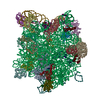
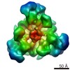
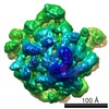
- PDB-3w1k: Crystal structure of the selenocysteine synthase SelA and tRNASec... -
+ Open data

- Basic information
| Entry | Database: PDB / ID: 3w1k | ||||||
|---|---|---|---|---|---|---|---|
| Title | Crystal structure of the selenocysteine synthase SelA and tRNASec complex | ||||||
 Components Components |
| ||||||
 Keywords Keywords | TRANSFERASE/RNA / protein-RNA complex / homodecamer / pentamer of dimers / fold-type I pyridoxal 5'-phosphate (PLP) dependent enzyme / non-canonical tRNA / L-seryl-tRNA(Sec) selenium transferase / selenocysteine synthesis / selenium metabolism / TRANSFERASE-RNA complex | ||||||
| Function / homology |  Function and homology information Function and homology informationL-seryl-tRNASec selenium transferase / L-seryl-tRNA(Sec) selenium transferase activity / selenocysteine incorporation / identical protein binding / cytoplasm Similarity search - Function | ||||||
| Biological species |   Aquifex aeolicus (bacteria)Thermoanaerobacter tengcongensis MB4 (bacteria) Aquifex aeolicus (bacteria)Thermoanaerobacter tengcongensis MB4 (bacteria) | ||||||
| Method |  X-RAY DIFFRACTION / SYNCHROTRON / MOLECULAR REPLACEMENT / Resolution: 7.5 Å X-RAY DIFFRACTION / SYNCHROTRON / MOLECULAR REPLACEMENT / Resolution: 7.5 Å | ||||||
 Authors Authors | Itoh, Y. / Sekine, S. / Yokoyama, S. | ||||||
 Citation Citation | Journal: Science / Year: 2013 Title: Decameric SelA-tRNA(Sec) ring structure reveals mechanism of bacterial selenocysteine formation Authors: Itoh, Y. / Brocker, M.J. / Sekine, S. / Hammond, G. / Suetsugu, S. / Soll, D. / Yokoyama, S. | ||||||
| History |
|
- Structure visualization
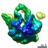
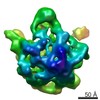
| Structure viewer | Molecule: MolmilJmol/JSmol |
|---|
- Downloads & links
-Download
| PDBx/mmCIF format | 3w1k.cif.gz | 694.1 KB | Display | PDBx/mmCIF format |
|---|---|---|---|---|
| PDB format | pdb3w1k.ent.gz | 561.2 KB | Display | PDB format |
| PDBx/mmJSON format | 3w1k.json.gz | Tree view | PDBx/mmJSON format | |
| Others |  Other downloads Other downloads |
-Validation report
| Summary document | 3w1k_validation.pdf.gz | 500.6 KB | Display | wwPDB validaton report |
|---|---|---|---|---|
| Full document | 3w1k_full_validation.pdf.gz | 651.8 KB | Display | |
| Data in XML | 3w1k_validation.xml.gz | 71 KB | Display | |
| Data in CIF | 3w1k_validation.cif.gz | 107.9 KB | Display | |
| Arichive directory | https://data.pdbj.org/pub/pdb/validation_reports/w1/3w1kftp://data.pdbj.org/pub/pdb/validation_reports/w1/3w1k | HTTPS FTP |
-Related structure data
| Related structure data | 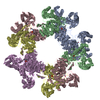 3w1hSC 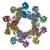 3w1iC  3w1jC S: Starting model for refinement C: citing same article ( |
|---|---|
| Similar structure data |

-Links
 PDBj
PDBj


- Assembly
| Deposited unit | 
| ||||||||
|---|---|---|---|---|---|---|---|---|---|
| 1 | 
| ||||||||
| Unit cell |
|
-Components
| #1: Protein | Mass: 50925.160 Da / Num. of mol.: 5 / Mutation: K19A, K21A, K46A, K48A Source method: isolated from a genetically manipulated source Source: (gene. exp.) Aquifex aeolicus (bacteria) / Strain: VF5 / Gene: selA / Plasmid: pET25b / Production host: References: UniProt: O67140, L-seryl-tRNASec selenium transferase #2: RNA chain | Mass: 30492.035 Da / Num. of mol.: 5 / Source method: obtained synthetically Details: The tRNASec from Thermoanaerobacter tengcongensis MB4 was prepared by in vitro transcription with T7 RNA polymerase. Source: (synth.) Thermoanaerobacter tengcongensis MB4 (bacteria)References: GenBank: 20671658 |
|---|
-Experimental details
-Experiment
| Experiment | Method: X-RAY DIFFRACTION / Number of used crystals: 1 |
|---|
- Sample preparation
| Crystal | Density Matthews: 4.85 Å3/Da / Density % sol: 74.66 % |
|---|---|
| Crystal grow | Temperature: 293 K / Method: vapor diffusion, sitting drop / pH: 7.5 Details: 35% ethylene glycol, 95mM NaNO3, 20mM Tris-HCl, 0.2M NaCl, 10mM MgCl2, 3mM 2-mercaptoethanol, pH 7.5, VAPOR DIFFUSION, SITTING DROP, temperature 293K |
-Data collection
| Diffraction | Mean temperature: 90 K |
|---|---|
| Diffraction source | Source: SYNCHROTRON / Site: Photon Factory  / Beamline: AR-NW12A / Wavelength: 1 Å / Beamline: AR-NW12A / Wavelength: 1 Å |
| Detector | Type: ADSC QUANTUM 210 / Detector: CCD / Date: Mar 16, 2009 / Details: mirrors |
| Radiation | Monochromator: Si / Protocol: SINGLE WAVELENGTH / Monochromatic (M) / Laue (L): M / Scattering type: x-ray |
| Radiation wavelength | Wavelength: 1 Å / Relative weight: 1 |
| Reflection | Resolution: 7.5→50 Å / Num. all: 9956 / Num. obs: 9777 / % possible obs: 98.2 % / Observed criterion σ(I): -1 / Redundancy: 3.6 % / Biso Wilson estimate: 250 Å2 / Rmerge(I) obs: 0.102 / Rsym value: 0.102 / Net I/σ(I): 16.3 |
| Reflection shell | Resolution: 7.5→7.77 Å / Redundancy: 2.9 % / Rmerge(I) obs: 0.712 / Mean I/σ(I) obs: 2.04 / Num. unique all: 990 / Rsym value: 0.712 / % possible all: 97.3 |
- Processing
| Software |
| ||||||||||||||||||||||||||||||||||||
|---|---|---|---|---|---|---|---|---|---|---|---|---|---|---|---|---|---|---|---|---|---|---|---|---|---|---|---|---|---|---|---|---|---|---|---|---|---|
| Refinement | Method to determine structure: MOLECULAR REPLACEMENT Starting model: 3W1H Resolution: 7.5→49.94 Å / Rfactor Rfree error: 0.011 / Data cutoff high absF: 7615012.16 / Data cutoff low absF: 0 / Isotropic thermal model: RESTRAINED / Cross valid method: THROUGHOUT / σ(F): 0 / Details: BULK SOLVENT MODEL USED
| ||||||||||||||||||||||||||||||||||||
| Solvent computation | Solvent model: FLAT MODEL / Bsol: 314.482 Å2 / ksol: 0.3 e/Å3 | ||||||||||||||||||||||||||||||||||||
| Displacement parameters | Biso mean: 525.5 Å2
| ||||||||||||||||||||||||||||||||||||
| Refine analyze |
| ||||||||||||||||||||||||||||||||||||
| Refinement step | Cycle: LAST / Resolution: 7.5→49.94 Å
| ||||||||||||||||||||||||||||||||||||
| Refine LS restraints |
| ||||||||||||||||||||||||||||||||||||
| LS refinement shell | Resolution: 7.5→7.97 Å / Rfactor Rfree error: 0.044 / Total num. of bins used: 6
| ||||||||||||||||||||||||||||||||||||
| Xplor file |
|
