negative regulation of SREBP signaling pathway / negative regulation of triglyceride biosynthetic process / regulation of cell migration involved in sprouting angiogenesis / negative regulation of hepatocyte proliferation / positive regulation of epidermal growth factor-activated receptor activity / regulation of lipid storage / Parkin-FBXW7-Cul1 ubiquitin ligase complex / ubiquitin-protein transferase activator activity / F-box domain binding / ubiquitin recycling ...negative regulation of SREBP signaling pathway / negative regulation of triglyceride biosynthetic process / regulation of cell migration involved in sprouting angiogenesis / negative regulation of hepatocyte proliferation / positive regulation of epidermal growth factor-activated receptor activity / regulation of lipid storage / Parkin-FBXW7-Cul1 ubiquitin ligase complex / ubiquitin-protein transferase activator activity / F-box domain binding / ubiquitin recycling / PcG protein complex / phosphothreonine residue binding / regulation of cell cycle G1/S phase transition / regulation of autophagy of mitochondrion / negative regulation of osteoclast development / positive regulation of oxidative stress-induced neuron intrinsic apoptotic signaling pathway / positive regulation of proteasomal protein catabolic process / Cul7-RING ubiquitin ligase complex / positive regulation of ubiquitin protein ligase activity / maintenance of protein location in nucleus / positive regulation of ubiquitin-dependent protein catabolic process / Loss of Function of FBXW7 in Cancer and NOTCH1 Signaling / vasculature development / positive regulation of ubiquitin-protein transferase activity / sister chromatid cohesion / SCF-dependent proteasomal ubiquitin-dependent protein catabolic process / positive regulation of protein targeting to mitochondrion / SCF ubiquitin ligase complex / ubiquitin ligase complex scaffold activity / negative regulation of Notch signaling pathway / protein monoubiquitination / Prolactin receptor signaling / Association of TriC/CCT with target proteins during biosynthesis / lipid homeostasis / cullin family protein binding / ubiquitin-like ligase-substrate adaptor activity / protein K48-linked ubiquitination / vasculogenesis / Nuclear events stimulated by ALK signaling in cancer / Notch signaling pathway / Regulation of BACH1 activity / cyclin binding / MAP3K8 (TPL2)-dependent MAPK1/3 activation / ubiquitin binding / positive regulation of protein ubiquitination / SCF-beta-TrCP mediated degradation of Emi1 / NIK-->noncanonical NF-kB signaling / molecular function activator activity / Vpu mediated degradation of CD4 / Dectin-1 mediated noncanonical NF-kB signaling / Degradation of GLI1 by the proteasome / Activation of NF-kappaB in B cells / lung development / Negative regulation of NOTCH4 signaling / Iron uptake and transport / GSK3B and BTRC:CUL1-mediated-degradation of NFE2L2 / Degradation of GLI2 by the proteasome / GLI3 is processed to GLI3R by the proteasome / FBXL7 down-regulates AURKA during mitotic entry and in early mitosis / protein destabilization / Degradation of beta-catenin by the destruction complex / regulation of circadian rhythm / NOTCH1 Intracellular Domain Regulates Transcription / CLEC7A (Dectin-1) signaling / beta-catenin binding / SCF(Skp2)-mediated degradation of p27/p21 / Constitutive Signaling by NOTCH1 PEST Domain Mutants / Constitutive Signaling by NOTCH1 HD+PEST Domain Mutants / FCERI mediated NF-kB activation / protein polyubiquitination / Interleukin-1 signaling / Orc1 removal from chromatin / Regulation of RUNX2 expression and activity / cellular response to UV / rhythmic process / Cyclin D associated events in G1 / regulation of protein localization / Regulation of PLK1 Activity at G2/M Transition / protein-macromolecule adaptor activity / Antigen processing: Ubiquitination & Proteasome degradation / Circadian Clock / Downstream TCR signaling / chromosome / Neddylation / proteasome-mediated ubiquitin-dependent protein catabolic process / positive regulation of ERK1 and ERK2 cascade / protein stabilization / protein ubiquitination / chromatin remodeling / protein domain specific binding / negative regulation of gene expression / DNA repair / centrosome / DNA damage response / ubiquitin protein ligase binding / nucleolus / perinuclear region of cytoplasm / Golgi apparatus / endoplasmic reticulum / protein-containing complex Similarity search - Function
In the structure databanks used in Yorodumi, some data are registered as the other names, "COVID-19 virus" and "2019-nCoV". Here are the details of the virus and the list of structure data.
Jan 31, 2019. EMDB accession codes are about to change! (news from PDBe EMDB page)
EMDB accession codes are about to change! (news from PDBe EMDB page)
The allocation of 4 digits for EMDB accession codes will soon come to an end. Whilst these codes will remain in use, new EMDB accession codes will include an additional digit and will expand incrementally as the available range of codes is exhausted. The current 4-digit format prefixed with “EMD-” (i.e. EMD-XXXX) will advance to a 5-digit format (i.e. EMD-XXXXX), and so on. It is currently estimated that the 4-digit codes will be depleted around Spring 2019, at which point the 5-digit format will come into force.
The EM Navigator/Yorodumi systems omit the EMD- prefix.
Related info.:Q: What is EMD? / ID/Accession-code notation in Yorodumi/EM Navigator
Yorodumi is a browser for structure data from EMDB, PDB, SASBDB, etc.
This page is also the successor to EM Navigator detail page, and also detail information page/front-end page for Omokage search.
The word "yorodu" (or yorozu) is an old Japanese word meaning "ten thousand". "mi" (miru) is to see.
Related info.:EMDB / PDB / SASBDB / Comparison of 3 databanks / Yorodumi Search / Aug 31, 2016. New EM Navigator & Yorodumi / Yorodumi Papers / Jmol/JSmol / Function and homology information / Changes in new EM Navigator and Yorodumi
 Movie
Movie Controller


 Open data
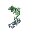
 Basic information
 Components
 Keywords
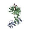
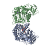
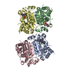
 F-box / WD40 domains / double phosphorylation / TRANSCRIPTION-CELL CYCLE COMPLEX
 Function and homology information
 Authors
 Citation
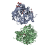
 Structure visualization
 Downloads & links
 Other downloads

 PDBj
PDBj


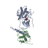
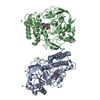
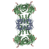
 Assembly


 Mass: 96.063 Da / Num. of mol.: 8 / Source method: obtained synthetically / Formula: SO4
 Mass: 18.015 Da / Num. of mol.: 178 / Source method: isolated from a natural source / Formula: H2O
 Sample preparation
 / Beamline: X9A / Wavelength: 0.9795 Å
 Processing