 Movie
Movie Controller


+ Open data

- Basic information
| Entry | Database: PDB / ID: 2o9u | ||||||
|---|---|---|---|---|---|---|---|
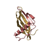
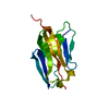
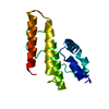
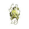
| Title | Monellin (MNEI) at 1.15 resolution | ||||||
 Components Components | Monellin chain B and Monellin chain A | ||||||
 Keywords Keywords |  PLANT PROTEIN / alternative conformations / 3-10 polyproline II helix PLANT PROTEIN / alternative conformations / 3-10 polyproline II helix | ||||||
| Function / homology |  Function and homology informationMonellin, A chain / Monellin, A chain superfamily / Monellin, B chain / Monellin / Monellin / Nuclear Transport Factor 2; Chain: A, - #10 / Cystatin superfamily / Nuclear Transport Factor 2; Chain: A, / Roll / Alpha Beta Function and homology informationMonellin, A chain / Monellin, A chain superfamily / Monellin, B chain / Monellin / Monellin / Nuclear Transport Factor 2; Chain: A, - #10 / Cystatin superfamily / Nuclear Transport Factor 2; Chain: A, / Roll / Alpha BetaSimilarity search - Domain/homology | ||||||
| Biological species |  Dioscoreophyllum cumminsii (serendipity berry) Dioscoreophyllum cumminsii (serendipity berry) | ||||||
| Method | X-RAY DIFFRACTION / SYNCHROTRON / MOLECULAR REPLACEMENT / Resolution: 1.15 Å | ||||||
 Authors Authors | Hobbs, J.R. / Munger, S.D. / Conn, G.L. | ||||||
 Citation Citation | Journal: Acta Crystallogr.,Sect.F / Year: 2007 Title: Monellin (MNEI) at 1.15 A resolution Authors: Hobbs, J.R. / Munger, S.D. / Conn, G.L. | ||||||
| History |
|
- Structure visualization
| Structure viewer | Molecule: MolmilJmol/JSmol |
|---|
- Downloads & links
-Download
| PDBx/mmCIF format | 2o9u.cif.gz | 72.4 KB | Display | PDBx/mmCIF format |
|---|---|---|---|---|
| PDB format | pdb2o9u.ent.gz | 54.4 KB | Display | PDB format |
| PDBx/mmJSON format | 2o9u.json.gz | Tree view | PDBx/mmJSON format | |
| Others |  Other downloads Other downloads |
-Validation report
| Arichive directory | https://data.pdbj.org/pub/pdb/validation_reports/o9/2o9uftp://data.pdbj.org/pub/pdb/validation_reports/o9/2o9u | HTTPS FTP |
|---|
-Related structure data
| Related structure data |  1n98 S: Starting model for refinement |
|---|---|
| Similar structure data |


-Links
 PDBj
PDBj
- Assembly
| Deposited unit | 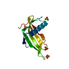
| ||||||||
|---|---|---|---|---|---|---|---|---|---|
| 1 |
| ||||||||
| Unit cell |
|
-Components
| #1: Protein | Mass: 11419.049 Da / Num. of mol.: 1 Source method: isolated from a genetically manipulated source Source: (gene. exp.) Dioscoreophyllum cumminsii (serendipity berry)Plasmid: pET22b / Production host:  Escherichia coli (E. coli) / References: UniProt: P02882, UniProt: P02881 Escherichia coli (E. coli) / References: UniProt: P02882, UniProt: P02881 | ||||
|---|---|---|---|---|---|
| #2: Chemical | ChemComp-SO4 / Sulfate  Mass: 96.063 Da / Num. of mol.: 4 / Source method: obtained synthetically / Formula: SO4 Mass: 96.063 Da / Num. of mol.: 4 / Source method: obtained synthetically / Formula: SO4#3: Water | ChemComp-HOH / | Water Mass: 18.015 Da / Num. of mol.: 142 / Source method: isolated from a natural source / Formula: H2O Mass: 18.015 Da / Num. of mol.: 142 / Source method: isolated from a natural source / Formula: H2OSequence details | REFER TO REF. 2 IN UNP DATABASE MONB_DIOCU(P02882). | |
-Experimental details
-Experiment
| Experiment | Method: X-RAY DIFFRACTION / Number of used crystals: 1 |
|---|
- Sample preparation
| Crystal | Density Matthews: 1.99 Å3/Da / Density % sol: 38.06 % |
|---|---|
| Crystal grow | Temperature: 291 K / Method: vapor diffusion, hanging drop / pH: 5.6 Details: 0.2M Ammonium sulfate, 30% PEG4000, pH 5.6, VAPOR DIFFUSION, HANGING DROP, temperature 291K |
-Data collection
| Diffraction | Mean temperature: 100 K |
|---|---|
| Diffraction source | Source: SYNCHROTRON / Site: ESRF  / Beamline: ID14-2 / Wavelength: 0.933 Å / Beamline: ID14-2 / Wavelength: 0.933 Å |
| Detector | Type: ADSC QUANTUM 4 / Detector: CCD / Date: Oct 1, 2004 |
| Radiation | Monochromator: Diamond(111), Ge(220) / Protocol: SINGLE WAVELENGTH / Monochromatic (M) / Laue (L): M / Scattering type: x-ray |
| Radiation wavelength | Wavelength: 0.933 Å / Relative weight: 1 |
| Reflection | Resolution: 1.15→22.44 Å / Num. obs: 27957 / % possible obs: 88.3 % / Observed criterion σ(F): 0 / Observed criterion σ(I): 0 / Redundancy: 3.8 % / Biso Wilson estimate: 9.4 Å2 / Rmerge(I) obs: 0.043 |
| Reflection shell | Resolution: 1.15→1.21 Å / Redundancy: 3.5 % / Rmerge(I) obs: 0.298 / Mean I/σ(I) obs: 4.5 / Num. unique all: 2513 / % possible all: 54.2 |
- Processing
| Software |
| |||||||||||||||||||||||||||||||||||||||||||||||||||||||||||||||||||||||||||||||||||||||||||||||||||||||||||||||||||||||||||||||||||||||||||||||||
|---|---|---|---|---|---|---|---|---|---|---|---|---|---|---|---|---|---|---|---|---|---|---|---|---|---|---|---|---|---|---|---|---|---|---|---|---|---|---|---|---|---|---|---|---|---|---|---|---|---|---|---|---|---|---|---|---|---|---|---|---|---|---|---|---|---|---|---|---|---|---|---|---|---|---|---|---|---|---|---|---|---|---|---|---|---|---|---|---|---|---|---|---|---|---|---|---|---|---|---|---|---|---|---|---|---|---|---|---|---|---|---|---|---|---|---|---|---|---|---|---|---|---|---|---|---|---|---|---|---|---|---|---|---|---|---|---|---|---|---|---|---|---|---|---|---|---|
| Refinement | Method to determine structure: MOLECULAR REPLACEMENT Starting model: 1N98 1n98 Resolution: 1.15→22.44 Å / Cor.coef. Fo:Fc: 0.977 / Cor.coef. Fo:Fc free: 0.969 / SU B: 1.234 / SU ML: 0.026 / Cross valid method: THROUGHOUT / σ(F): 0 / ESU R: 0.044 / ESU R Free: 0.043 / Stereochemistry target values: MAXIMUM LIKELIHOOD / Details: HYDROGENS HAVE BEEN ADDED IN THE RIDING POSITIONS
| |||||||||||||||||||||||||||||||||||||||||||||||||||||||||||||||||||||||||||||||||||||||||||||||||||||||||||||||||||||||||||||||||||||||||||||||||
| Solvent computation | Ion probe radii: 0.8 Å / Shrinkage radii: 0.8 Å / VDW probe radii: 1.2 Å / Solvent model: BABINET MODEL WITH MASK | |||||||||||||||||||||||||||||||||||||||||||||||||||||||||||||||||||||||||||||||||||||||||||||||||||||||||||||||||||||||||||||||||||||||||||||||||
| Displacement parameters | Biso mean: 12.723 Å2
| |||||||||||||||||||||||||||||||||||||||||||||||||||||||||||||||||||||||||||||||||||||||||||||||||||||||||||||||||||||||||||||||||||||||||||||||||
| Refinement step | Cycle: LAST / Resolution: 1.15→22.44 Å
| |||||||||||||||||||||||||||||||||||||||||||||||||||||||||||||||||||||||||||||||||||||||||||||||||||||||||||||||||||||||||||||||||||||||||||||||||
| Refine LS restraints |
| |||||||||||||||||||||||||||||||||||||||||||||||||||||||||||||||||||||||||||||||||||||||||||||||||||||||||||||||||||||||||||||||||||||||||||||||||
| LS refinement shell | Resolution: 1.15→1.18 Å / Total num. of bins used: 20
|
