 Movie
Movie Controller

[English] 日本語
 Yorodumi
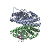
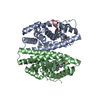
- PDB-1qkm: HUMAN OESTROGEN RECEPTOR BETA LIGAND-BINDING DOMAIN IN COMPLEX WI... -
+ Open data

- Basic information
| Entry | Database: PDB / ID: 1qkm | ||||||
|---|---|---|---|---|---|---|---|
| Title | HUMAN OESTROGEN RECEPTOR BETA LIGAND-BINDING DOMAIN IN COMPLEX WITH PARTIAL AGONIST GENISTEIN | ||||||
 Components Components | ESTROGEN RECEPTOR BETA | ||||||
 Keywords Keywords | NUCLEAR RECEPTOR / TRANSCRIPTION FACTOR / PHYTO-OESTROGEN / PARTIAL AGONIST | ||||||
| Function / homology |  Function and homology informationreceptor antagonist activity / nuclear steroid receptor activity / nuclear estrogen receptor activity / intracellular estrogen receptor signaling pathway / cellular response to estrogen stimulus / estrogen response element binding / steroid binding / ESR-mediated signaling / cellular response to estradiol stimulus / negative regulation of cell growth ...receptor antagonist activity / nuclear steroid receptor activity / nuclear estrogen receptor activity / intracellular estrogen receptor signaling pathway / cellular response to estrogen stimulus / estrogen response element binding / steroid binding / ESR-mediated signaling / cellular response to estradiol stimulus / negative regulation of cell growth / Nuclear Receptor transcription pathway / Constitutive Signaling by Aberrant PI3K in Cancer / nuclear receptor activity / positive regulation of DNA-binding transcription factor activity / cell-cell signaling / PIP3 activates AKT signaling / PI5P, PP2A and IER3 Regulate PI3K/AKT Signaling / Extra-nuclear estrogen signaling / DNA-binding transcription factor activity, RNA polymerase II-specific / RNA polymerase II cis-regulatory region sequence-specific DNA binding / intracellular membrane-bounded organelle / chromatin / regulation of DNA-templated transcription / regulation of transcription by RNA polymerase II / positive regulation of DNA-templated transcription / negative regulation of transcription by RNA polymerase II / enzyme binding / signal transduction / positive regulation of transcription by RNA polymerase II / mitochondrion / DNA binding / zinc ion binding / nucleoplasm / nucleus Function and homology informationreceptor antagonist activity / nuclear steroid receptor activity / nuclear estrogen receptor activity / intracellular estrogen receptor signaling pathway / cellular response to estrogen stimulus / estrogen response element binding / steroid binding / ESR-mediated signaling / cellular response to estradiol stimulus / negative regulation of cell growth ...receptor antagonist activity / nuclear steroid receptor activity / nuclear estrogen receptor activity / intracellular estrogen receptor signaling pathway / cellular response to estrogen stimulus / estrogen response element binding / steroid binding / ESR-mediated signaling / cellular response to estradiol stimulus / negative regulation of cell growth / Nuclear Receptor transcription pathway / Constitutive Signaling by Aberrant PI3K in Cancer / nuclear receptor activity / positive regulation of DNA-binding transcription factor activity / cell-cell signaling / PIP3 activates AKT signaling / PI5P, PP2A and IER3 Regulate PI3K/AKT Signaling / Extra-nuclear estrogen signaling / DNA-binding transcription factor activity, RNA polymerase II-specific / RNA polymerase II cis-regulatory region sequence-specific DNA binding / intracellular membrane-bounded organelle / chromatin / regulation of DNA-templated transcription / regulation of transcription by RNA polymerase II / positive regulation of DNA-templated transcription / negative regulation of transcription by RNA polymerase II / enzyme binding / signal transduction / positive regulation of transcription by RNA polymerase II / mitochondrion / DNA binding / zinc ion binding / nucleoplasm / nucleusSimilarity search - Function | ||||||
| Biological species |  HOMO SAPIENS (human) HOMO SAPIENS (human) | ||||||
| Method | X-RAY DIFFRACTION / SYNCHROTRON / MOLECULAR REPLACEMENT / Resolution: 1.8 Å | ||||||
 Authors Authors | Pike, A.C.W. / Brzozowski, A.M. / Carlquist, M. | ||||||
 Citation Citation | Journal: Embo J. / Year: 1999 Title: Structure of the Ligand-Binding Domain of Oestrogen Receptor Beta in the Presence of a Partial Agonist and a Full Antagonist Authors: Pike, A.C.W. / Brzozowski, A.M. / Hubbard, R.E. / Bonn, T. / Thorsell, A.-G. / Engstrom, O. / Ljunggren, J. / Gustaffson, J.-A. / Carlquist, M. #1: Journal: Nature / Year: 1997Title: Molecular Basis of Agonism and Antagonism in the Oestrogen Receptor Authors: Brzozowski, A.M. / Pike, A.C.W. / Dauter, Z. / Hubbard, R.E. / Bonn, T. / Engstrom, O. / Ohman, L. / Greene, G.L. / Gustaffson, J.-A. / Carlquist, M. | ||||||
| History |
|
- Structure visualization
| Structure viewer | Molecule: MolmilJmol/JSmol |
|---|
- Downloads & links
-Download
| PDBx/mmCIF format | 1qkm.cif.gz | 64 KB | Display | PDBx/mmCIF format |
|---|---|---|---|---|
| PDB format | pdb1qkm.ent.gz | 46.5 KB | Display | PDB format |
| PDBx/mmJSON format | 1qkm.json.gz | Tree view | PDBx/mmJSON format | |
| Others |  Other downloads Other downloads |
-Validation report
| Arichive directory | https://data.pdbj.org/pub/pdb/validation_reports/qk/1qkmftp://data.pdbj.org/pub/pdb/validation_reports/qk/1qkm | HTTPS FTP |
|---|
-Related structure data
| Related structure data |  1qknC 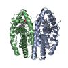 1ereS C: citing same article ( S: Starting model for refinement |
|---|---|
| Similar structure data |


-Links
 PDBj
PDBj
- Assembly
| Deposited unit | 
| ||||||||
|---|---|---|---|---|---|---|---|---|---|
| 1 | 
| ||||||||
| Unit cell |
| ||||||||
| Details | BIOLOGICAL_UNIT: DIMER |
-Components
| #1: Protein | / OESTROGEN RECEPTOR / ER-LBD Mass: 28726.092 Da / Num. of mol.: 1 / Fragment: LIGAND-BINDING DOMAIN Source method: isolated from a genetically manipulated source Details: COMPLEXED WITH THE PHYTO-OESTROGEN GENISTEIN / Source: (gene. exp.) HOMO SAPIENS (human) / Gene: OESTROGEN RECEPTOR BETA / Plasmid: PLEX / Production host:  ESCHERICHIA COLI (E. coli) / Strain (production host): GI724 / References: UniProt: Q92731 ESCHERICHIA COLI (E. coli) / Strain (production host): GI724 / References: UniProt: Q92731 |
|---|---|
| #2: Chemical | ChemComp-GEN / Genistein  Mass: 270.237 Da / Num. of mol.: 1 / Source method: obtained synthetically / Formula: C15H10O5 / Comment: inhibitor*YM Mass: 270.237 Da / Num. of mol.: 1 / Source method: obtained synthetically / Formula: C15H10O5 / Comment: inhibitor*YM |
| #3: Water | ChemComp-HOH / Water Mass: 18.015 Da / Num. of mol.: 139 / Source method: isolated from a natural source / Formula: H2O Mass: 18.015 Da / Num. of mol.: 139 / Source method: isolated from a natural source / Formula: H2O |
-Experimental details
-Experiment
| Experiment | Method: X-RAY DIFFRACTION / Number of used crystals: 1 |
|---|
- Sample preparation
| Crystal | Density Matthews: 2.44 Å3/Da / Density % sol: 49 % | ||||||||||||||||||||||||||||||
|---|---|---|---|---|---|---|---|---|---|---|---|---|---|---|---|---|---|---|---|---|---|---|---|---|---|---|---|---|---|---|---|
| Crystal grow | pH: 8.1 Details: 6-9% (W/V) PEG 6000, 1.6-2.1M NACL, 0.1M TRIS-HCL, PH 8.1 | ||||||||||||||||||||||||||||||
| Crystal grow | *PLUS Method: vapor diffusion, hanging dropDetails: drop consists of equal volume of protein and reservoir solutions | ||||||||||||||||||||||||||||||
| Components of the solutions | *PLUS
|
-Data collection
| Diffraction | Mean temperature: 100 K |
|---|---|
| Diffraction source | Source: SYNCHROTRON / Site: ESRF  / Beamline: ID14-4 / Wavelength: 0.93 / Beamline: ID14-4 / Wavelength: 0.93 |
| Detector | Type: ADSC CCD / Detector: CCD / Date: Dec 15, 1998 |
| Radiation | Protocol: SINGLE WAVELENGTH / Monochromatic (M) / Laue (L): M / Scattering type: x-ray |
| Radiation wavelength | Wavelength: 0.93 Å / Relative weight: 1 |
| Reflection | Resolution: 1.8→60 Å / Num. obs: 28523 / % possible obs: 99.7 % / Observed criterion σ(I): -3 / Redundancy: 8 % / Biso Wilson estimate: 32.6 Å2 / Rsym value: 0.049 / Net I/σ(I): 15 |
| Reflection shell | Resolution: 1.8→1.83 Å / Redundancy: 8 % / Mean I/σ(I) obs: 4 / Rsym value: 0.433 / % possible all: 100 |
| Reflection | *PLUS Num. measured all: 358818 / Rmerge(I) obs: 0.049 |
| Reflection shell | *PLUS % possible obs: 100 % / Rmerge(I) obs: 0.433 |
- Processing
| Software |
| ||||||||||||||||||||||||||||||||||||||||||||||||||||||||||||||||||||||||||||||||||||
|---|---|---|---|---|---|---|---|---|---|---|---|---|---|---|---|---|---|---|---|---|---|---|---|---|---|---|---|---|---|---|---|---|---|---|---|---|---|---|---|---|---|---|---|---|---|---|---|---|---|---|---|---|---|---|---|---|---|---|---|---|---|---|---|---|---|---|---|---|---|---|---|---|---|---|---|---|---|---|---|---|---|---|---|---|---|
| Refinement | Method to determine structure: MOLECULAR REPLACEMENT Starting model: PDB ENTRY 1ERE Resolution: 1.8→55 Å / Cross valid method: THROUGHOUT / σ(F): 0 / ESU R: 0.12715 / ESU R Free: 0.12556 Details: BULK SOLVENT CORRECTION CALCULATED IN XPLOR V3.843 WAS USED THROUGHOUT. DISCREPANCY BETWEEN REFINEMENT STATISTICS GIVEN ABOVE AND PUBLISHED VALUES ARISE DUE TO A SEQUENCE ERROR THAT HAS ...Details: BULK SOLVENT CORRECTION CALCULATED IN XPLOR V3.843 WAS USED THROUGHOUT. DISCREPANCY BETWEEN REFINEMENT STATISTICS GIVEN ABOVE AND PUBLISHED VALUES ARISE DUE TO A SEQUENCE ERROR THAT HAS SUBSEQUENTLY BEEN CORRECTED.
| ||||||||||||||||||||||||||||||||||||||||||||||||||||||||||||||||||||||||||||||||||||
| Displacement parameters | Biso mean: 41.3 Å2
| ||||||||||||||||||||||||||||||||||||||||||||||||||||||||||||||||||||||||||||||||||||
| Refinement step | Cycle: LAST / Resolution: 1.8→55 Å
| ||||||||||||||||||||||||||||||||||||||||||||||||||||||||||||||||||||||||||||||||||||
| Refine LS restraints |
|
