 Movie
Movie Controller


+ Open data

- Basic information
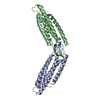
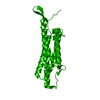
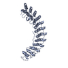
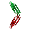
| Entry | Database: PDB / ID: 1n11 | ||||||
|---|---|---|---|---|---|---|---|
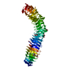
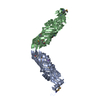
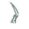
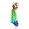
| Title | D34 REGION OF HUMAN ANKYRIN-R AND LINKER | ||||||
 Components Components | Ankyrin | ||||||
 Keywords Keywords | STRUCTURAL PROTEIN / ANKYRIN / CLATHRIN / BAND 3 / ANION EXCHANGER | ||||||
| Function / homology |  Function and homology information Function and homology informationpositive regulation of organelle organization / spectrin-associated cytoskeleton / maintenance of epithelial cell apical/basal polarity / NrCAM interactions / ankyrin-1 complex / Neurofascin interactions / CHL1 interactions / cytoskeletal anchor activity / M band / Interaction between L1 and Ankyrins ...positive regulation of organelle organization / spectrin-associated cytoskeleton / maintenance of epithelial cell apical/basal polarity / NrCAM interactions / ankyrin-1 complex / Neurofascin interactions / CHL1 interactions / cytoskeletal anchor activity / M band / Interaction between L1 and Ankyrins / spectrin binding / exocytosis / axolemma / endoplasmic reticulum to Golgi vesicle-mediated transport / COPI-mediated anterograde transport / cytoskeleton organization / sarcoplasmic reticulum / protein localization to plasma membrane / sarcolemma / structural constituent of cytoskeleton / cytoplasmic side of plasma membrane / Z disc / ATPase binding / postsynaptic membrane / basolateral plasma membrane / protein phosphatase binding / transmembrane transporter binding / cytoskeleton / neuron projection / structural molecule activity / enzyme binding / signal transduction / plasma membrane / cytosolSimilarity search - Function | ||||||
| Biological species |  Homo sapiens (human) Homo sapiens (human) | ||||||
| Method | X-RAY DIFFRACTION / SYNCHROTRON / MAD / Resolution: 2.7 Å | ||||||
 Authors Authors | Michaely, P. / Tomchick, D.R. / Machius, M. / Anderson, R.G.W. | ||||||
 Citation Citation | Journal: Embo J. / Year: 2002 Title: Crystal structure of a 12 ANK repeat stack from human ankyrinR Authors: Michaely, P. / Tomchick, D.R. / Machius, M. / Anderson, R.G.W. | ||||||
| History |
|
- Structure visualization
| Structure viewer | Molecule: MolmilJmol/JSmol |
|---|
- Downloads & links
-Download
| PDBx/mmCIF format | 1n11.cif.gz | 87.9 KB | Display | PDBx/mmCIF format |
|---|---|---|---|---|
| PDB format | pdb1n11.ent.gz | 67.1 KB | Display | PDB format |
| PDBx/mmJSON format | 1n11.json.gz | Tree view | PDBx/mmJSON format | |
| Others |  Other downloads Other downloads |
-Validation report
| Arichive directory | https://data.pdbj.org/pub/pdb/validation_reports/n1/1n11ftp://data.pdbj.org/pub/pdb/validation_reports/n1/1n11 | HTTPS FTP |
|---|
-Related structure data
| Similar structure data |
|---|


-Links
 PDBj
PDBj


- Assembly
| Deposited unit | 
| ||||||||
|---|---|---|---|---|---|---|---|---|---|
| 1 |
| ||||||||
| Unit cell |
|
-Components
| #1: Protein | Mass: 46459.898 Da / Num. of mol.: 1 / Fragment: D34 region Source method: isolated from a genetically manipulated source Source: (gene. exp.) Homo sapiens (human) / Gene: ank-1 / Plasmid: pGEX-kg / Production host:  Escherichia coli (E. coli) / Strain (production host): B834 / References: UniProt: P16157 Escherichia coli (E. coli) / Strain (production host): B834 / References: UniProt: P16157 |
|---|---|
| #2: Chemical | ChemComp-BR / Bromide  Mass: 79.904 Da / Num. of mol.: 1 / Source method: obtained synthetically / Formula: Br Mass: 79.904 Da / Num. of mol.: 1 / Source method: obtained synthetically / Formula: Br |
| #3: Chemical | ChemComp-CL / Chloride  Mass: 35.453 Da / Num. of mol.: 9 / Source method: obtained synthetically / Formula: Cl Mass: 35.453 Da / Num. of mol.: 9 / Source method: obtained synthetically / Formula: Cl |
-Experimental details
-Experiment
| Experiment | Method: X-RAY DIFFRACTION / Number of used crystals: 1 |
|---|
- Sample preparation
| Crystal grow | Temperature: 277 K / Method: vapor diffusion, hanging drop / pH: 7.5 Details: PEG400, HEPES, NaBr, CaCl2, acetonitrile, pH 7.5, VAPOR DIFFUSION, HANGING DROP, temperature 277K | ||||||||||||||||||||||||||||||||||||||||||||||||||||||||||||||||||||||
|---|---|---|---|---|---|---|---|---|---|---|---|---|---|---|---|---|---|---|---|---|---|---|---|---|---|---|---|---|---|---|---|---|---|---|---|---|---|---|---|---|---|---|---|---|---|---|---|---|---|---|---|---|---|---|---|---|---|---|---|---|---|---|---|---|---|---|---|---|---|---|---|
| Crystal grow | *PLUS Temperature: 4 ℃ / Method: vapor diffusion | ||||||||||||||||||||||||||||||||||||||||||||||||||||||||||||||||||||||
| Components of the solutions | *PLUS
|
-Data collection
| Diffraction | Mean temperature: 100 K |
|---|---|
| Diffraction source | Source: SYNCHROTRON / Site: APS  / Beamline: 19-ID / Wavelength: 0.97925 Å / Beamline: 19-ID / Wavelength: 0.97925 Å |
| Detector | Type: SBC-2 / Detector: CCD / Date: Jan 31, 2001 |
| Radiation | Monochromator: Graphite Double Crystal / Protocol: SINGLE WAVELENGTH / Monochromatic (M) / Laue (L): M / Scattering type: x-ray |
| Radiation wavelength | Wavelength: 0.97925 Å / Relative weight: 1 |
| Reflection | Resolution: 2.5→50 Å / Num. all: 421780 / Num. obs: 415031 / % possible obs: 98.4 % / Observed criterion σ(F): 0 / Observed criterion σ(I): 0 / Biso Wilson estimate: 67 Å2 / Rmerge(I) obs: 0.082 / Net I/σ(I): 22.6 |
| Reflection shell | Resolution: 2.5→2.54 Å / Rmerge(I) obs: 0.699 / Mean I/σ(I) obs: 1.3 / % possible all: 93 |
| Reflection | *PLUS Lowest resolution: 50 Å / Num. obs: 37994 / Num. measured all: 415031 |
| Reflection shell | *PLUS % possible obs: 93 % |
- Processing
| Software |
| |||||||||||||||||||||
|---|---|---|---|---|---|---|---|---|---|---|---|---|---|---|---|---|---|---|---|---|---|---|
| Refinement | Method to determine structure: MAD / Resolution: 2.7→30 Å / Isotropic thermal model: individual isotropic / Cross valid method: THROUGHOUT / σ(F): 0 / Stereochemistry target values: Engh & Huber
| |||||||||||||||||||||
| Displacement parameters | Biso mean: 67 Å2
| |||||||||||||||||||||
| Refine analyze | Luzzati sigma a obs: 1.07 Å | |||||||||||||||||||||
| Refinement step | Cycle: LAST / Resolution: 2.7→30 Å
| |||||||||||||||||||||
| Refine LS restraints |
| |||||||||||||||||||||
| Refinement | *PLUS Lowest resolution: 30 Å / % reflection Rfree: 3.7 % | |||||||||||||||||||||
| Solvent computation | *PLUS | |||||||||||||||||||||
| Displacement parameters | *PLUS | |||||||||||||||||||||
| Refine LS restraints | *PLUS Type: c_bond_d / Dev ideal: 0.01 |
