 Movie
Movie Controller

[English] 日本語
 Yorodumi
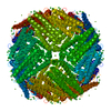
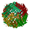
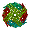
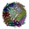
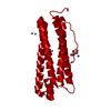
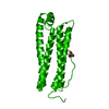
- PDB-1fha: SOLVING THE STRUCTURE OF HUMAN H FERRITIN BY GENETICALLY ENGINEER... -
+ Open data

- Basic information
| Entry | Database: PDB / ID: 1fha | ||||||
|---|---|---|---|---|---|---|---|
| Title | SOLVING THE STRUCTURE OF HUMAN H FERRITIN BY GENETICALLY ENGINEERING INTERMOLECULAR CRYSTAL CONTACTS | ||||||
 Components Components | FERRITIN | ||||||
 Keywords Keywords | METAL BINDING PROTEIN / IRON STORAGE | ||||||
| Function / homology |  Function and homology information Function and homology informationiron ion sequestering activity / Scavenging by Class A Receptors / intracellular ferritin complex / autolysosome / Golgi Associated Vesicle Biogenesis / ferroxidase / intracellular sequestering of iron ion / ferroxidase activity / negative regulation of fibroblast proliferation / ferric iron binding ...iron ion sequestering activity / Scavenging by Class A Receptors / intracellular ferritin complex / autolysosome / Golgi Associated Vesicle Biogenesis / ferroxidase / intracellular sequestering of iron ion / ferroxidase activity / negative regulation of fibroblast proliferation / ferric iron binding / ferrous iron binding / Iron uptake and transport / tertiary granule lumen / iron ion transport / intracellular iron ion homeostasis / ficolin-1-rich granule lumen / immune response / iron ion binding / negative regulation of cell population proliferation / Neutrophil degranulation / extracellular exosome / extracellular region / identical protein binding / nucleus / cytosol / cytoplasmSimilarity search - Function | ||||||
| Biological species |  Homo sapiens (human) Homo sapiens (human) | ||||||
| Method | X-RAY DIFFRACTION / Resolution: 2.4 Å | ||||||
 Authors Authors | Artymiuk, P.J. / Harrison, P.M. | ||||||
 Citation Citation | Journal: Nature / Year: 1991 Title: Solving the structure of human H ferritin by genetically engineering intermolecular crystal contacts. Authors: Lawson, D.M. / Artymiuk, P.J. / Yewdall, S.J. / Smith, J.M. / Livingstone, J.C. / Treffry, A. / Luzzago, A. / Levi, S. / Arosio, P. / Cesareni, G. / Thomas, C.D. / Shaw, W.V. / Harrison, P.M. | ||||||
| History |
| ||||||
| Remark 700 | SHEET THERE IS A STRAND FROM 84 TO 86 THAT FORMS A TENUOUS TWO-STRANDED ANIT-PARALLEL BETA SHEET ...SHEET THERE IS A STRAND FROM 84 TO 86 THAT FORMS A TENUOUS TWO-STRANDED ANIT-PARALLEL BETA SHEET WITH THE EQUIVALENT STRAND IN A TWO-FOLD RELATED SUBUNIT. |
- Structure visualization
| Structure viewer | Molecule: MolmilJmol/JSmol |
|---|
- Downloads & links
-Download
| PDBx/mmCIF format | 1fha.cif.gz | 47.6 KB | Display | PDBx/mmCIF format |
|---|---|---|---|---|
| PDB format | pdb1fha.ent.gz | 34.9 KB | Display | PDB format |
| PDBx/mmJSON format | 1fha.json.gz | Tree view | PDBx/mmJSON format | |
| Others |  Other downloads Other downloads |
-Validation report
| Arichive directory | https://data.pdbj.org/pub/pdb/validation_reports/fh/1fhaftp://data.pdbj.org/pub/pdb/validation_reports/fh/1fha | HTTPS FTP |
|---|
-Related structure data
| Similar structure data |
|---|


-Links
 PDBj
PDBj


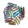
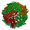
- Assembly
| Deposited unit | 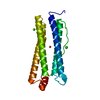
| |||||||||
|---|---|---|---|---|---|---|---|---|---|---|
| 1 | x 24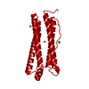
| |||||||||
| Unit cell |
| |||||||||
| Atom site foot note | 1: RESIDUE 161 IS A CIS PROLINE. | |||||||||
| Components on special symmetry positions |
|
-Components
| #1: Protein | Mass: 21254.605 Da / Num. of mol.: 1 Source method: isolated from a genetically manipulated source Source: (gene. exp.) Homo sapiens (human) / References: UniProt: P02794 | ||||||
|---|---|---|---|---|---|---|---|
| #2: Chemical | ChemComp-FE / Iron  Mass: 55.845 Da / Num. of mol.: 1 / Source method: obtained synthetically / Formula: Fe Mass: 55.845 Da / Num. of mol.: 1 / Source method: obtained synthetically / Formula: Fe | ||||||
| #3: Chemical |   Mass: 40.078 Da / Num. of mol.: 2 / Source method: obtained synthetically / Formula: Ca Mass: 40.078 Da / Num. of mol.: 2 / Source method: obtained synthetically / Formula: Ca#4: Water | ChemComp-HOH / | Water Mass: 18.015 Da / Num. of mol.: 20 / Source method: isolated from a natural source / Formula: H2O Mass: 18.015 Da / Num. of mol.: 20 / Source method: isolated from a natural source / Formula: H2OCompound details | THE FERROXIDASE CENTER WHICH CATALYSES THE OXIDATION OF FE(II) TO FE(III) INVOLVES THE FOLLOWING ...THE FERROXIDAS | Sequence details | THE FERRITIN SEQUENCE CLONED IN ESCHERICHIA COLI WAS THAT OF HOMO SAPIENS EXCEPT THAT LYS 86 WAS ...THE FERRITIN SEQUENCE CLONED IN ESCHERICHI | |
-Experimental details
-Experiment
| Experiment | Method: X-RAY DIFFRACTION |
|---|
- Sample preparation
| Crystal | Density Matthews: 3.09 Å3/Da / Density % sol: 60.23 % |
|---|
-Data collection
| Radiation | Scattering type: x-ray |
|---|---|
| Radiation wavelength | Relative weight: 1 |
| Reflection | *PLUS Highest resolution: 2.4 Å / Lowest resolution: 20 Å / Num. obs: 10318 / % possible obs: 95 % / Num. measured all: 54965 / Rmerge(I) obs: 0.128 |
- Processing
| Software | Name: PROLSQ / Classification: refinement | ||||||||||||||||||||||||||||||||||||||||||||||||||||||||||||||||||||||||||||||||||||
|---|---|---|---|---|---|---|---|---|---|---|---|---|---|---|---|---|---|---|---|---|---|---|---|---|---|---|---|---|---|---|---|---|---|---|---|---|---|---|---|---|---|---|---|---|---|---|---|---|---|---|---|---|---|---|---|---|---|---|---|---|---|---|---|---|---|---|---|---|---|---|---|---|---|---|---|---|---|---|---|---|---|---|---|---|---|
| Refinement | Rfactor obs: 0.205 / Highest resolution: 2.4 Å Details: THE STRUCTURE WAS SOLVED BY GENETICALLY ENGINEERING INTERMOLECULAR CRYSTAL CONTACTS FROM THE KNOWN STRUCTURE OF HORSE L CHAIN FERRITIN INTO HUMAN H CHAIN FERRITIN. | ||||||||||||||||||||||||||||||||||||||||||||||||||||||||||||||||||||||||||||||||||||
| Refinement step | Cycle: LAST / Highest resolution: 2.4 Å
| ||||||||||||||||||||||||||||||||||||||||||||||||||||||||||||||||||||||||||||||||||||
| Refine LS restraints |
| ||||||||||||||||||||||||||||||||||||||||||||||||||||||||||||||||||||||||||||||||||||
| Refinement | *PLUS Lowest resolution: 20 Å / Rfactor obs: 0.205 | ||||||||||||||||||||||||||||||||||||||||||||||||||||||||||||||||||||||||||||||||||||
| Solvent computation | *PLUS | ||||||||||||||||||||||||||||||||||||||||||||||||||||||||||||||||||||||||||||||||||||
| Displacement parameters | *PLUS |
