 Movie
Movie Controller

[English] 日本語
 Yorodumi
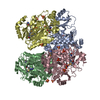
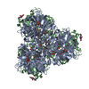
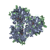
- PDB-1e3p: tungstate derivative of Streptomyces antibioticus PNPase/GPSI enzyme -
+ Open data

- Basic information
| Entry | Database: PDB / ID: 1e3p | ||||||
|---|---|---|---|---|---|---|---|
| Title | tungstate derivative of Streptomyces antibioticus PNPase/GPSI enzyme | ||||||
 Components Components | Polyribonucleotide nucleotidyltransferase Polynucleotide phosphorylase Polynucleotide phosphorylase | ||||||
 Keywords Keywords | POLYRIBONUCLEOTIDE TRANSFERASE / ATP-GTP DIPHOSPHOTRANSFERASE RNA PROCESSING / RNA DEGRADATION | ||||||
| Function / homology |  Function and homology informationpolyribonucleotide nucleotidyltransferase / polyribonucleotide nucleotidyltransferase activity / mRNA catabolic process / RNA processing / magnesium ion binding / RNA binding / cytoplasm Function and homology informationpolyribonucleotide nucleotidyltransferase / polyribonucleotide nucleotidyltransferase activity / mRNA catabolic process / RNA processing / magnesium ion binding / RNA binding / cytoplasmSimilarity search - Function | ||||||
| Biological species |  Streptomyces antibioticus (bacteria) Streptomyces antibioticus (bacteria) | ||||||
| Method | X-RAY DIFFRACTION / MOLECULAR REPLACEMENT / Resolution: 2.5 Å | ||||||
 Authors Authors | Symmons, M.F. / Jones, G.H. / Luisi, B.F. | ||||||
 Citation Citation | Journal: Structure / Year: 2000 Title: A Duplicated Fold is the Structural Basis for Polynucleotide Phosphorylase Catalytic Activity, Processivity, and Regulation Authors: Symmons, M.F. / Jones, G.H. / Luisi, B.F. | ||||||
| History |
|
- Structure visualization
| Structure viewer | Molecule: MolmilJmol/JSmol |
|---|
- Downloads & links
-Download
| PDBx/mmCIF format | 1e3p.cif.gz | 143.1 KB | Display | PDBx/mmCIF format |
|---|---|---|---|---|
| PDB format | pdb1e3p.ent.gz | 108 KB | Display | PDB format |
| PDBx/mmJSON format | 1e3p.json.gz | Tree view | PDBx/mmJSON format | |
| Others |  Other downloads Other downloads |
-Validation report
| Arichive directory | https://data.pdbj.org/pub/pdb/validation_reports/e3/1e3pftp://data.pdbj.org/pub/pdb/validation_reports/e3/1e3p | HTTPS FTP |
|---|
-Related structure data
| Related structure data |  1e3hSC S: Starting model for refinement C: citing same article ( |
|---|---|
| Similar structure data |


-Links
 PDBj
PDBj


- Assembly
| Deposited unit | 
| ||||||||
|---|---|---|---|---|---|---|---|---|---|
| 1 | 
| ||||||||
| Unit cell |
| ||||||||
| Details | ENZYME IS TRIMER IN SOLUTION |
-Components
| #1: Protein | Polynucleotide phosphorylase / Polynucleotide phosphorylase / PNPase Mass: 81225.367 Da / Num. of mol.: 1 Source method: isolated from a genetically manipulated source Details: BIFUNCTIONAL ENZYME POLYRIBONUCLEOTIDE NUCLEOTIDYL TRANSFERASE, ATP-GTP DIPHOSPHOTRANSFERASE Source: (gene. exp.) Streptomyces antibioticus (bacteria) / Description: BIFUNCTIONAL ENZYME ISOLATED / Cellular location: CYTOPLASM / Gene: pnp, AFM16_28085 / Cellular location (production host): CYTOPLASM / Production host: ESCHERICHIA COLI (E. coli)References: UniProt: A0A1S9NJJ0, UniProt: Q53597*PLUS, polyribonucleotide nucleotidyltransferase | ||||||
|---|---|---|---|---|---|---|---|
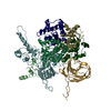
| #2: Chemical | ChemComp-SO4 / Sulfate  Mass: 96.063 Da / Num. of mol.: 10 / Source method: obtained synthetically / Formula: SO4 Mass: 96.063 Da / Num. of mol.: 10 / Source method: obtained synthetically / Formula: SO4#3: Chemical | ChemComp-WO4 / |   Mass: 247.838 Da / Num. of mol.: 1 / Source method: obtained synthetically / Formula: WO4 Mass: 247.838 Da / Num. of mol.: 1 / Source method: obtained synthetically / Formula: WO4#4: Water | ChemComp-HOH / | Water Mass: 18.015 Da / Num. of mol.: 305 / Source method: isolated from a natural source / Formula: H2O Mass: 18.015 Da / Num. of mol.: 305 / Source method: isolated from a natural source / Formula: H2OSequence details | ARG A 31, SEQUENCING AMBIGUITY ILE A 156, SEQUENCING AMBIGUITY ILE A 210, SEQUENCING AMBIGUITY PHE ...ARG A 31, SEQUENCING | |
-Experimental details
-Experiment
| Experiment | Method: X-RAY DIFFRACTION / Number of used crystals: 1 |
|---|
- Sample preparation
| Crystal | Density Matthews: 3.33 Å3/Da / Density % sol: 54 % Description: SELENOMETHIONINES WERE REPLACED WITH METHIONINES FOR MOLECULAR REPLACEMENT | ||||||||||||||||||||||||||||||||||||||||||
|---|---|---|---|---|---|---|---|---|---|---|---|---|---|---|---|---|---|---|---|---|---|---|---|---|---|---|---|---|---|---|---|---|---|---|---|---|---|---|---|---|---|---|---|
| Crystal grow | pH: 7 Details: 2.0M (NH4)2SO4, 100MM TRISHCL PH8.5, 100MM BISTRISHCL PH6.5, 60MM NACL, 4MM MGCL2, 5MM DTT, 50MM NA2W04, pH 7.00 | ||||||||||||||||||||||||||||||||||||||||||
| Crystal grow | *PLUS Temperature: 20 ℃ / pH: 8.5 / Method: vapor diffusion | ||||||||||||||||||||||||||||||||||||||||||
| Components of the solutions | *PLUS
|
-Data collection
| Diffraction | Mean temperature: 100 K |
|---|---|
| Diffraction source | Source: ROTATING ANODE / Type: MSC / Wavelength: 1.5418 |
| Detector | Type: R-AXIS IV / Detector: IMAGE PLATE / Date: Apr 15, 1998 / Details: YALE MIRRORS |
| Radiation | Protocol: SINGLE WAVELENGTH / Monochromatic (M) / Laue (L): M / Scattering type: x-ray |
| Radiation wavelength | Wavelength: 1.5418 Å / Relative weight: 1 |
| Reflection | Resolution: 2.5→20 Å / Num. obs: 37642 / % possible obs: 99.3 % / Redundancy: 6.5 % / Biso Wilson estimate: 59.1 Å2 / Rsym value: 0.079 / Net I/σ(I): 17.7 |
| Reflection shell | Resolution: 2.5→2.56 Å / Redundancy: 2.8 % / Mean I/σ(I) obs: 2.8 / Rsym value: 0.38 / % possible all: 92.3 |
| Reflection | *PLUS Num. measured all: 462572 / Rmerge(I) obs: 0.08 |
- Processing
| Software |
| ||||||||||||||||||||||||||||||||||||||||||||||||||||||||||||||||||||||||||||||||
|---|---|---|---|---|---|---|---|---|---|---|---|---|---|---|---|---|---|---|---|---|---|---|---|---|---|---|---|---|---|---|---|---|---|---|---|---|---|---|---|---|---|---|---|---|---|---|---|---|---|---|---|---|---|---|---|---|---|---|---|---|---|---|---|---|---|---|---|---|---|---|---|---|---|---|---|---|---|---|---|---|---|
| Refinement | Method to determine structure: MOLECULAR REPLACEMENT Starting model: 1E3H Resolution: 2.5→19.84 Å / Rfactor Rfree error: 0.004 / Data cutoff high absF: 5669653.7 / Isotropic thermal model: RESTRAINED / Cross valid method: THROUGHOUT / σ(F): 0 Details: REFINEMENT TARGET (MLF) INCLUDED ANOMALOUS DATA POOR DENSITY FOR RESIDUES 604 - 614 AND 623 - 634 WAS INTERPRETED FROM STRUCTURE OF HOMOLOGOUS DOMAIN PDB 1VIH. POOR DENSITY FOR RESIDUES 656 - ...Details: REFINEMENT TARGET (MLF) INCLUDED ANOMALOUS DATA POOR DENSITY FOR RESIDUES 604 - 614 AND 623 - 634 WAS INTERPRETED FROM STRUCTURE OF HOMOLOGOUS DOMAIN PDB 1VIH. POOR DENSITY FOR RESIDUES 656 - 661, 663 - 671, 675 - 679, AND 699 - 717 WAS INTERPRETED FROM STRUCTURE OF HOMOLOGOUS DOMAIN PDB 1SRO. MODEL HERE IS POLYALA (EXCEPT GLY AND PRO WHERE EXPECTED FROM SEQUENCE) WITH B-FACTOR SET TO 100.00 AND SUBJECT TO POSITIONAL REFINEMENT ONLY. AFTER POSITIONAL REFINEMENT RMSD CA ATOMS WERE 1.4 A (OVER 28 EQUIVALENT ATOMS) AND 1.6 (OVER 39 EQUIVALENT ATOMS) FOR 1VIH AND 1SRO HOMOLOGOUS DOMAINS RESPECTIVELY. THE C-TERMINAL RESIDUE WAS NOT SEEN IN ELECTRON DENSITY MAP
| ||||||||||||||||||||||||||||||||||||||||||||||||||||||||||||||||||||||||||||||||
| Solvent computation | Solvent model: FLAT MODEL / Bsol: 52.0576 Å2 / ksol: 0.339268 e/Å3 | ||||||||||||||||||||||||||||||||||||||||||||||||||||||||||||||||||||||||||||||||
| Displacement parameters | Biso mean: 50.6 Å2
| ||||||||||||||||||||||||||||||||||||||||||||||||||||||||||||||||||||||||||||||||
| Refine analyze |
| ||||||||||||||||||||||||||||||||||||||||||||||||||||||||||||||||||||||||||||||||
| Refinement step | Cycle: LAST / Resolution: 2.5→19.84 Å
| ||||||||||||||||||||||||||||||||||||||||||||||||||||||||||||||||||||||||||||||||
| Refine LS restraints |
| ||||||||||||||||||||||||||||||||||||||||||||||||||||||||||||||||||||||||||||||||
| LS refinement shell | Resolution: 2.5→2.66 Å / Rfactor Rfree error: 0.016 / Total num. of bins used: 6
| ||||||||||||||||||||||||||||||||||||||||||||||||||||||||||||||||||||||||||||||||
| Xplor file |
| ||||||||||||||||||||||||||||||||||||||||||||||||||||||||||||||||||||||||||||||||
| Software | *PLUS Name: CNS / Version: 0.9A / Classification: refinement | ||||||||||||||||||||||||||||||||||||||||||||||||||||||||||||||||||||||||||||||||
| Refine LS restraints | *PLUS
|
