 Movie
Movie Controller


+ Open data

- Basic information
| Entry | Database: PDB / ID: 1cqe | |||||||||
|---|---|---|---|---|---|---|---|---|---|---|
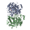
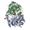
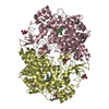
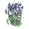
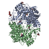
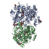
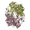
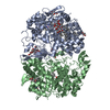
| Title | PROSTAGLANDIN H2 SYNTHASE-1 COMPLEX WITH FLURBIPROFEN | |||||||||
 Components Components | PROTEIN (PROSTAGLANDIN H2 SYNTHASE-1) | |||||||||
 Keywords Keywords | OXIDOREDUCTASE / DIOXYGENASE / PEROXIDASE | |||||||||
| Function / homology |  Function and homology information Function and homology informationprostaglandin-endoperoxide synthase / prostaglandin-endoperoxide synthase activity / oxidoreductase activity, acting on single donors with incorporation of molecular oxygen, incorporation of two atoms of oxygen / cyclooxygenase pathway / prostaglandin biosynthetic process / regulation of blood pressure / peroxidase activity / response to oxidative stress / neuron projection / intracellular membrane-bounded organelle ...prostaglandin-endoperoxide synthase / prostaglandin-endoperoxide synthase activity / oxidoreductase activity, acting on single donors with incorporation of molecular oxygen, incorporation of two atoms of oxygen / cyclooxygenase pathway / prostaglandin biosynthetic process / regulation of blood pressure / peroxidase activity / response to oxidative stress / neuron projection / intracellular membrane-bounded organelle / endoplasmic reticulum membrane / heme binding / protein homodimerization activity / metal ion binding / cytoplasm Similarity search - Function | |||||||||
| Biological species |  | |||||||||
| Method |  X-RAY DIFFRACTION / MIR / Resolution: 3.1 Å X-RAY DIFFRACTION / MIR / Resolution: 3.1 Å | |||||||||
 Authors Authors | Picot, D. / Loll, P.J. / Mulichak, A.M. / Garavito, R.M. | |||||||||
 Citation Citation | Journal: Nature / Year: 1994 Title: The X-ray crystal structure of the membrane protein prostaglandin H2 synthase-1. Authors: Picot, D. / Loll, P.J. / Garavito, R.M. #1: Journal: Biochemistry / Year: 1996Title: Synthesis and Use of Iodinated Nonsteroidal Antiinflammatory Drug Analogs as Crystallographic Probes of the Prostaglandin H2 Synthase Cyclooxygenase Active Site Authors: Loll, P.J. / Picot, D. / Ekabo, O. / Garavito, R.M. #2: Journal: Nat.Struct.Biol. / Year: 1995Title: The Structural Basis of Aspirin Activity Inferred from the Crystal Structure of Inactivated Prostaglandin H2 Synthase Authors: Loll, P.J. / Picot, D. / Garavito, R.M. #3: Journal: J.Mol.Biol. / Year: 1992Title: X-Ray Crystal Structure of Canine Myeloperoxidase at 3 Angstroms Resolution Authors: Zeng, J. / Fenna, R.E. | |||||||||
| History |
|
- Structure visualization
| Structure viewer | Molecule: MolmilJmol/JSmol |
|---|
- Downloads & links
-Download
| PDBx/mmCIF format | 1cqe.cif.gz | 243.5 KB | Display | PDBx/mmCIF format |
|---|---|---|---|---|
| PDB format | pdb1cqe.ent.gz | 196.4 KB | Display | PDB format |
| PDBx/mmJSON format | 1cqe.json.gz | Tree view | PDBx/mmJSON format | |
| Others |  Other downloads Other downloads |
-Validation report
| Summary document | 1cqe_validation.pdf.gz | 1023.7 KB | Display | wwPDB validaton report |
|---|---|---|---|---|
| Full document | 1cqe_full_validation.pdf.gz | 1 MB | Display | |
| Data in XML | 1cqe_validation.xml.gz | 27.1 KB | Display | |
| Data in CIF | 1cqe_validation.cif.gz | 40.6 KB | Display | |
| Arichive directory | https://data.pdbj.org/pub/pdb/validation_reports/cq/1cqeftp://data.pdbj.org/pub/pdb/validation_reports/cq/1cqe | HTTPS FTP |
-Related structure data


-Links
 PDBj
PDBj


- Assembly
| Deposited unit | 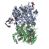
| ||||||||||
|---|---|---|---|---|---|---|---|---|---|---|---|
| 1 |
| ||||||||||
| Unit cell |
| ||||||||||
| Noncrystallographic symmetry (NCS) | NCS oper: (Code: given Matrix: (-0.99514, -0.06069, 0.0775), Vector: |
-Components
-Protein , 1 types, 2 molecules AB
| #1: Protein | Mass: 66595.320 Da / Num. of mol.: 2 / Source method: isolated from a natural source Details: PROTEIN IS GLYCOSYLATED AT RESIDUES ASN68, ASN144 AND ASN410 Source: (natural) References: UniProt: P05979, prostaglandin-endoperoxide synthase |
|---|
-Sugars , 2 types, 11 molecules 
| #2: Polysaccharide | 2-acetamido-2-deoxy-beta-D-glucopyranose-(1-4)-2-acetamido-2-deoxy-beta-D-glucopyranose Source method: isolated from a genetically manipulated source #3: Sugar | ChemComp-BOG /  Type: D-saccharide / Mass: 292.369 Da / Num. of mol.: 5 / Source method: obtained synthetically / Formula: C14H28O6 / Comment: detergent*YM Type: D-saccharide / Mass: 292.369 Da / Num. of mol.: 5 / Source method: obtained synthetically / Formula: C14H28O6 / Comment: detergent*YM |
|---|
-Non-polymers , 3 types, 134 molecules 

| #4: Chemical |  Mass: 616.487 Da / Num. of mol.: 2 / Source method: obtained synthetically / Formula: C34H32FeN4O4 Mass: 616.487 Da / Num. of mol.: 2 / Source method: obtained synthetically / Formula: C34H32FeN4O4#5: Chemical |  Mass: 244.261 Da / Num. of mol.: 2 / Source method: obtained synthetically / Formula: C15H13FO2 / Comment: antiinflammatory*YM Mass: 244.261 Da / Num. of mol.: 2 / Source method: obtained synthetically / Formula: C15H13FO2 / Comment: antiinflammatory*YM#6: Water | ChemComp-HOH / | Mass: 18.015 Da / Num. of mol.: 130 / Source method: isolated from a natural source / Formula: H2O |
|---|
-Experimental details
-Experiment
| Experiment | Method: X-RAY DIFFRACTION / Number of used crystals: 4 |
|---|
- Sample preparation
| Crystal | Density Matthews: 2.9 Å3/Da / Density % sol: 73.08 % | ||||||||||||||||||||||||||||||||||||||||||||||||
|---|---|---|---|---|---|---|---|---|---|---|---|---|---|---|---|---|---|---|---|---|---|---|---|---|---|---|---|---|---|---|---|---|---|---|---|---|---|---|---|---|---|---|---|---|---|---|---|---|---|
| Crystal grow | Method: vapor diffusion, hanging drop / pH: 6.7 Details: CRYSTALLIZED BY HANGING DROP VAPOR DIFFUSION METHOD USING 1:1 RATIO OF PROTEIN SOLUTION (10 MG/ML IN 20 MM SODIUM PHOSPHATE BUFFER PH6.7, 100-200 MM NACL, 0.6% BETA-OCTYL GLUCOPYRANOSIDE, 0. ...Details: CRYSTALLIZED BY HANGING DROP VAPOR DIFFUSION METHOD USING 1:1 RATIO OF PROTEIN SOLUTION (10 MG/ML IN 20 MM SODIUM PHOSPHATE BUFFER PH6.7, 100-200 MM NACL, 0.6% BETA-OCTYL GLUCOPYRANOSIDE, 0.1 MM FLURBIPORFEN) AND RESERVOIR OF 4-8% PEG 4000., VAPOR DIFFUSION, HANGING DROP | ||||||||||||||||||||||||||||||||||||||||||||||||
| Crystal grow | *PLUS | ||||||||||||||||||||||||||||||||||||||||||||||||
| Components of the solutions | *PLUS
|
-Data collection
| Diffraction | Mean temperature: 277 K |
|---|---|
| Diffraction source | Source: ROTATING ANODE / Type: ELLIOTT GX-21 / Wavelength: 1.5418 |
| Detector | Detector: AREA DETECTOR |
| Radiation | Monochromator: GRAPHITE / Protocol: SINGLE WAVELENGTH / Monochromatic (M) / Laue (L): M / Scattering type: x-ray |
| Radiation wavelength | Wavelength: 1.5418 Å / Relative weight: 1 |
| Reflection | Resolution: 3.1→20 Å / Num. obs: 32349 / % possible obs: 96 % / Observed criterion σ(I): 2 / Rmerge(I) obs: 0.076 |
| Reflection shell | Resolution: 3.1→3.2 Å |
| Reflection | *PLUS Num. measured all: 112745 |
- Processing
| Software |
| ||||||||||||||||||||||||||||||||||||||||||||||||||||||||||||
|---|---|---|---|---|---|---|---|---|---|---|---|---|---|---|---|---|---|---|---|---|---|---|---|---|---|---|---|---|---|---|---|---|---|---|---|---|---|---|---|---|---|---|---|---|---|---|---|---|---|---|---|---|---|---|---|---|---|---|---|---|---|
| Refinement | Method to determine structure: MIR / Resolution: 3.1→20 Å / Cross valid method: THROUGHOUT / σ(F): 2
| ||||||||||||||||||||||||||||||||||||||||||||||||||||||||||||
| Refinement step | Cycle: LAST / Resolution: 3.1→20 Å
| ||||||||||||||||||||||||||||||||||||||||||||||||||||||||||||
| Refine LS restraints |
| ||||||||||||||||||||||||||||||||||||||||||||||||||||||||||||
| LS refinement shell | Resolution: 3.1→3.2 Å / Total num. of bins used: 8
| ||||||||||||||||||||||||||||||||||||||||||||||||||||||||||||
| Xplor file |
| ||||||||||||||||||||||||||||||||||||||||||||||||||||||||||||
| Software | *PLUS Name: X-PLOR / Version: 3.1 / Classification: refinement | ||||||||||||||||||||||||||||||||||||||||||||||||||||||||||||
| Refine LS restraints | *PLUS
|
