- EMDB-1285: Structure of the ribosome-bound cricket paralysis virus IRES RNA. -
+
データを開く
IDまたはキーワード:
読み込み中...
-
基本情報
登録情報
データベース: EMDB / ID: EMD-1285
タイトル
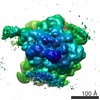
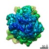
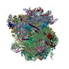
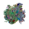
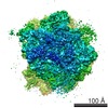
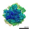
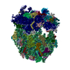
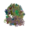
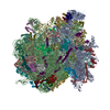
Structure of the ribosome-bound cricket paralysis virus IRES RNA.
マップデータ
Complex between the yeast 80S ribosome and the cricket paralysis virus IRES
試料
試料: yeast 80S ribosome in complex with the CrPV IRES RNAEukaryotic ribosome
複合体: yeast 80S ribosomeEukaryotic ribosome
RNA: CrPV IRESCripavirus internal ribosome entry site
機能・相同性
機能・相同性情報
: / Formation of the ternary complex, and subsequently, the 43S complex / Translation initiation complex formation / Ribosomal scanning and start codon recognition / Major pathway of rRNA processing in the nucleolus and cytosol / SRP-dependent cotranslational protein targeting to membrane / 90S preribosome / GTP hydrolysis and joining of the 60S ribosomal subunit / Formation of a pool of free 40S subunits / Nonsense Mediated Decay (NMD) independent of the Exon Junction Complex (EJC) ...: / Formation of the ternary complex, and subsequently, the 43S complex / Translation initiation complex formation / Ribosomal scanning and start codon recognition / Major pathway of rRNA processing in the nucleolus and cytosol / SRP-dependent cotranslational protein targeting to membrane / 90S preribosome / GTP hydrolysis and joining of the 60S ribosomal subunit / Formation of a pool of free 40S subunits / Nonsense Mediated Decay (NMD) independent of the Exon Junction Complex (EJC) / Nonsense Mediated Decay (NMD) enhanced by the Exon Junction Complex (EJC) / L13a-mediated translational silencing of Ceruloplasmin expression / ribosomal large subunit export from nucleus / regulation of translational fidelity / maturation of LSU-rRNA / ribosomal large subunit assembly / ribosomal small subunit assembly / cytosolic small ribosomal subunit / cytoplasmic translation / cytosolic large ribosomal subunit / rRNA binding / リボソーム / structural constituent of ribosome / 翻訳 (生物学) / mRNA binding / RNA binding / 細胞核 / 細胞質基質 / 細胞質 類似検索 - 分子機能
Ribosomal protein L1, conserved site / Ribosomal protein L1 / Ribosomal protein S5/S7, eukaryotic/archaeal / Ribosomal protein L1 signature. / Ribosomal protein L1, 3-layer alpha/beta-sandwich / Ribosomal protein L1-like / Ribosomal protein L1/ribosomal biogenesis protein / Ribosomal protein L1p/L10e family / Ribosomal protein L5, conserved site / Ribosomal protein S7, conserved site ...Ribosomal protein L1, conserved site / Ribosomal protein L1 / Ribosomal protein S5/S7, eukaryotic/archaeal / Ribosomal protein L1 signature. / Ribosomal protein L1, 3-layer alpha/beta-sandwich / Ribosomal protein L1-like / Ribosomal protein L1/ribosomal biogenesis protein / Ribosomal protein L1p/L10e family / Ribosomal protein L5, conserved site / Ribosomal protein S7, conserved site / Ribosomal protein L5, N-terminal / Ribosomal protein L5 signature. / Ribosomal protein L5, C-terminal / Ribosomal protein L5 / Ribosomal protein L5 domain superfamily / Ribosomal protein L5 / ribosomal L5P family C-terminus / Ribosomal protein S7 signature. / Ribosomal protein S5/S7 / Ribosomal protein S7 domain / Ribosomal protein S7 domain superfamily / Ribosomal protein S7p/S5e 類似検索 - ドメイン・相同性
Large ribosomal subunit protein uL1A / Small ribosomal subunit protein uS7 / 60S ribosomal protein L1-B / Large ribosomal subunit protein uL5B 類似検索 - 構成要素
ジャーナル: Nat Struct Mol Biol / 年: 2006 タイトル: Structure of the ribosome-bound cricket paralysis virus IRES RNA. 著者: Martin Schüler / Sean R Connell / Aurelie Lescoute / Jan Giesebrecht / Marylena Dabrowski / Birgit Schroeer / Thorsten Mielke / Pawel A Penczek / Eric Westhof / Christian M T Spahn / 要旨: Internal ribosome entry sites (IRESs) facilitate an alternative, end-independent pathway of translation initiation. A particular family of dicistroviral IRESs can assemble elongation-competent 80S ...Internal ribosome entry sites (IRESs) facilitate an alternative, end-independent pathway of translation initiation. A particular family of dicistroviral IRESs can assemble elongation-competent 80S ribosomal complexes in the absence of canonical initiation factors and initiator transfer RNA. We present here a cryo-EM reconstruction of a dicistroviral IRES bound to the 80S ribosome. The resolution of the cryo-EM reconstruction, in the subnanometer range, allowed the molecular structure of the complete IRES in its active, ribosome-bound state to be solved. The structure, harboring three pseudoknot-containing domains, each with a specific functional role, shows how defined elements of the IRES emerge from a compactly folded core and interact with the key ribosomal components that form the A, P and E sites, where tRNAs normally bind. Our results exemplify the molecular strategy for recruitment of an IRES and reveal the dynamic features necessary for internal initiation.
 ムービー
ムービー コントローラー


 データを開く
 基本情報
基本情報 マップデータ
 試料
試料 機能・相同性情報
 ribosomal large subunit export from nucleus / regulation of translational fidelity / maturation of LSU-rRNA /
 データ登録者
 引用
引用
 構造の表示
構造の表示
 ダウンロードとリンク
 1285.gif
 http://ftp.pdbj.org/pub/emdb/structures/EMD-1285
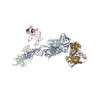


 試料の構成要素
 解析
解析 電子顕微鏡法
