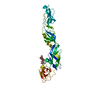
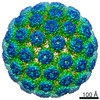
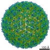
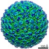
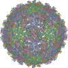
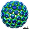
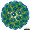
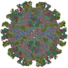
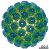
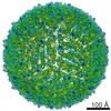
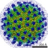
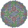
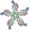
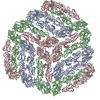
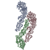
ジャーナル: J Struct Biol / 年: 2013 タイトル: Membrane curvature in flaviviruses. 著者: Wei Zhang / Bärbel Kaufmann / Paul R Chipman / Richard J Kuhn / Michael G Rossmann / 要旨: Coordinated interplay between membrane proteins and the lipid bilayer is required for such processes as transporter function and the entrance of enveloped viruses into host cells. In this study, ...Coordinated interplay between membrane proteins and the lipid bilayer is required for such processes as transporter function and the entrance of enveloped viruses into host cells. In this study, three-dimensional cryo-electron microscopy density maps of mature and immature flaviviruses were analyzed to assess the curvature of the membrane leaflets and its relation to membrane-bound viral glycoproteins. The overall morphology of the viral membrane is determined by the icosahedral scaffold composed of envelope (E) and membrane (M) proteins through interaction of the proteins' stem-anchor regions with the membrane. In localized regions, small membrane areas exhibit convex, concave, flat or saddle-shaped surfaces that are constrained by the specific protein organization within each membrane leaflet. These results suggest that the organization of membrane proteins in small enveloped viruses mediate the formation of membrane curvature.
名称: TNE (12 mM Tris, 120 mM NaCl, 1 mM EDTA) / pH: 8 / 詳細: 12 mM Tris-HCL, 120 mM NaCl, 1 mM EDTA
試料
濃度: 1 mg/ml / 包埋: NO / シャドウイング: NO / 染色: NO / 凍結: YES / 詳細: 12 mM Tris-HCL, 120 mM NaCl, 1 mM EDTA
試料支持
詳細: 400 mesh holey carbon copper grid
急速凍結
装置: HOMEMADE PLUNGER / 凍結剤: ETHANE / Temp: 98 K / 詳細: vitrification carried out in a BSL3 lab 手法: A small vial of ethane is placed inside a larger liquid nitrogen reservoir. The grid holding a few microliters of the sample is held in place at the bottom of a plunger by means of fine ...手法: A small vial of ethane is placed inside a larger liquid nitrogen reservoir. The grid holding a few microliters of the sample is held in place at the bottom of a plunger by means of fine tweezers. Once the ethane in the vial is completely frozen, it needs to be melted slightly. When the liquid ethane is ready, a piece of filter paper is then pressed against the sample to blot off excess buffer sufficient to leave a thin layer on the grid. After a predetermined time, the filter paper is removed and the plunger is allowed to drop into the liquid ethane. Once the grid enters the liquid ethane, the sample is rapidly frozen and the grid is transferred under liquid nitrogen to a storage box immersed in liquid nitrogen for later use in the microscope.
 ムービー
ムービー コントローラー


 データを開く
 基本情報
基本情報 要素
要素 キーワード
キーワード VIRUS (ウイルス) /
  機能・相同性情報
 データ登録者
 引用
引用
 構造の表示
構造の表示 ダウンロードとリンク
 その他のダウンロード
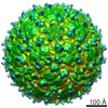
 PDBj
PDBj


 集合体
集合体
 試料調製
試料調製 電子顕微鏡撮影
 解析
解析