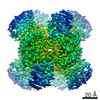
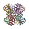
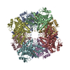
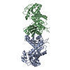
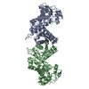
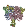
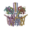
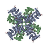
ジャーナル: J Struct Biol / 年: 2019 タイトル: Structural insights into thermostabilization of leucine dehydrogenase from its atomic structure by cryo-electron microscopy. 著者: Hiroki Yamaguchi / Akiko Kamegawa / Kunio Nakata / Tatsuki Kashiwagi / Toshimi Mizukoshi / Yoshinori Fujiyoshi / Kazutoshi Tani / 要旨: Leucine dehydrogenase (LDH, EC 1.4.1.9) is a NAD-dependent oxidoreductase that catalyzes the deamination of branched-chain l-amino acids (BCAAs). LDH of Geobacillus stearothermophilus (GstLDH) is a ...Leucine dehydrogenase (LDH, EC 1.4.1.9) is a NAD-dependent oxidoreductase that catalyzes the deamination of branched-chain l-amino acids (BCAAs). LDH of Geobacillus stearothermophilus (GstLDH) is a highly thermostable enzyme that has been applied for the quantification or production of BCAAs. Here the cryo-electron microscopy (cryo-EM) structures of apo and NAD-bound LDH are reported at 3.0 and 3.2 Å resolution, respectively. On comparing the structures, the two overall structures are almost identical, but it was observed that the partial conformational change was triggered by the interaction between Ser147 and the nicotinamide moiety of NAD. NAD binding also enhanced the strength of oligomerization interfaces formed by the core domains. Such additional interdomain interaction is in good agreement with our experimental results showing that the residual activity of NAD-bound form was approximately three times higher than that of the apo form after incubation at 80 °C. In addition, sequence comparison of three structurally known LDHs indicated a set of candidates for site-directed mutagenesis to improve thermostability. Subsequent mutation analysis actually revealed that non-conserved residues, including Ala94, Tyr127, and the C-terminal region, are crucial for oligomeric thermostability.
 ムービー
ムービー コントローラー


 データを開く
 基本情報
基本情報 要素
要素 ロイシンデヒドロゲナーゼ
  キーワード
キーワード 機能・相同性情報

 データ登録者
 日本, 1件
  引用
引用 構造の表示
構造の表示 ダウンロードとリンク
 その他のダウンロード
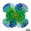
 PDBj
PDBj 集合体
集合体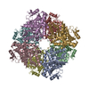
 試料調製
試料調製 電子顕微鏡撮影
 解析
解析