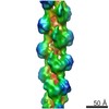
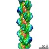
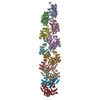
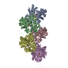
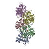
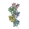
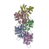
ジャーナル: Structure / 年: 2015 タイトル: Near-atomic resolution for one state of F-actin. 著者: Vitold E Galkin / Albina Orlova / Matthijn R Vos / Gunnar F Schröder / Edward H Egelman / 要旨: Actin functions as a helical polymer, F-actin, but attempts to build an atomic model for this filament have been hampered by the fact that the filament cannot be crystallized and by structural ...Actin functions as a helical polymer, F-actin, but attempts to build an atomic model for this filament have been hampered by the fact that the filament cannot be crystallized and by structural heterogeneity. We have used a direct electron detector, cryo-electron microscopy, and the forces imposed on actin filaments in thin films to reconstruct one state of the filament at 4.7 Å resolution, which allows for building a reliable pseudo-atomic model of F-actin. We also report a different state of the filament where actin protomers adopt a conformation observed in the crystal structure of the G-actin-profilin complex with an open ATP-binding cleft. Comparison of the two structural states provides insights into ATP-hydrolysis and filament dynamics. The atomic model provides a framework for understanding why every buried residue in actin has been under intense selective pressure.
らせん対称: (回転対称性: 1 / Dyad axis: no / N subunits divisor: 1 / Num. of operations: 5 / Rise per n subunits: 27.6 Å / Rotation per n subunits: -166.7 °)
詳細
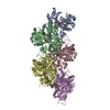
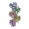
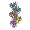
Actin forms a helical filament of indeterminate length. The designation "pentameric" in REMARK 350 is an artifact of the PDB format and can be disregarded.
 ムービー
ムービー コントローラー


 データを開く
 基本情報
基本情報 要素
要素
 キーワード
キーワード 機能・相同性情報
 データ登録者
 引用
引用


 構造の表示
構造の表示 ダウンロードとリンク
 その他のダウンロード
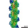
 PDBj
PDBj


 集合体
集合体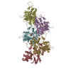

 分子量: 427.201 Da / 分子数: 5 / 由来タイプ: 合成 / 式: C10H15N5O10P2 / コメント: ADP, エネルギー貯蔵分子*YM
 分子量: 24.305 Da / 分子数: 5 / 由来タイプ: 合成 / 式: Mg
 試料調製
試料調製 電子顕微鏡撮影
 解析
解析