 ムービー
ムービー コントローラー


+ データを開く

- 基本情報
基本情報
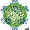
| 登録情報 | データベース: PDB / ID: 3j40 | |||||||||
|---|---|---|---|---|---|---|---|---|---|---|
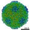
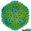
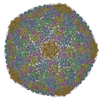
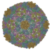
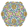
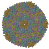
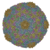
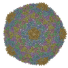
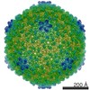
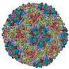
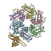
| タイトル | Validated Near-Atomic Resolution Structure of Bacteriophage Epsilon15 Derived from Cryo-EM and Modeling | |||||||||
 要素 要素 |
| |||||||||
 キーワード キーワード |  VIRUS (ウイルス) / capsid (カプシド) / accessory protein VIRUS (ウイルス) / capsid (カプシド) / accessory protein | |||||||||
| 機能・相同性 | viral capsid, decoration / カプシド / Major coat protein / Major capsid protein 機能・相同性情報 機能・相同性情報 | |||||||||
| 生物種 |  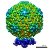 Salmonella phage epsilon15 (ファージ) Salmonella phage epsilon15 (ファージ) | |||||||||
| 手法 | 電子顕微鏡法 / 単粒子再構成法 / クライオ電子顕微鏡法 / 解像度: 4.5 Å | |||||||||
 データ登録者 データ登録者 | Baker, M.L. / Hryc, C.F. / Zhang, Q. / Wu, W. / Jakana, J. / Haase-Pettingell, C. / Afonine, P.V. / Adams, P.D. / King, J.A. / Jiang, W. / Chiu, W. | |||||||||
 引用 引用 | ジャーナル: Proc Natl Acad Sci U S A / 年: 2013 タイトル: Validated near-atomic resolution structure of bacteriophage epsilon15 derived from cryo-EM and modeling. 著者: Matthew L Baker / Corey F Hryc / Qinfen Zhang / Weimin Wu / Joanita Jakana / Cameron Haase-Pettingell / Pavel V Afonine / Paul D Adams / Jonathan A King / Wen Jiang / Wah Chiu /  要旨: High-resolution structures of viruses have made important contributions to modern structural biology. Bacteriophages, the most diverse and abundant organisms on earth, replicate and infect all ...High-resolution structures of viruses have made important contributions to modern structural biology. Bacteriophages, the most diverse and abundant organisms on earth, replicate and infect all bacteria and archaea, making them excellent potential alternatives to antibiotics and therapies for multidrug-resistant bacteria. Here, we improved upon our previous electron cryomicroscopy structure of Salmonella bacteriophage epsilon15, achieving a resolution sufficient to determine the tertiary structures of both gp7 and gp10 protein subunits that form the T = 7 icosahedral lattice. This study utilizes recently established best practice for near-atomic to high-resolution (3-5 Å) electron cryomicroscopy data evaluation. The resolution and reliability of the density map were cross-validated by multiple reconstructions from truly independent data sets, whereas the models of the individual protein subunits were validated adopting the best practices from X-ray crystallography. Some sidechain densities are clearly resolved and show the subunit-subunit interactions within and across the capsomeres that are required to stabilize the virus. The presence of the canonical phage and jellyroll viral protein folds, gp7 and gp10, respectively, in the same virus suggests that epsilon15 may have emerged more recently relative to other bacteriophages. | |||||||||
| 履歴 |
|
- 構造の表示
構造の表示
| ムービー |
ムービービューア |
|---|---|
| 構造ビューア | 分子: MolmilJmol/JSmol |
- ダウンロードとリンク
-ダウンロード
| PDBx/mmCIF形式 | 3j40.cif.gz | 605.6 KB | 表示 | PDBx/mmCIF形式 |
|---|---|---|---|---|
| PDB形式 | pdb3j40.ent.gz | 498.2 KB | 表示 | PDB形式 |
| PDBx/mmJSON形式 | 3j40.json.gz | ツリー表示 | PDBx/mmJSON形式 | |
| その他 |  その他のダウンロード その他のダウンロード |
-検証レポート
| アーカイブディレクトリ | https://data.pdbj.org/pub/pdb/validation_reports/j4/3j40ftp://data.pdbj.org/pub/pdb/validation_reports/j4/3j40 | HTTPS FTP |
|---|
-関連構造データ
-リンク
 PDBj
PDBj- 集合体
集合体
| 登録構造単位 | 
|
|---|---|
| 1 | x 60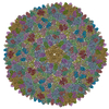
|
| 2 |
|
| 3 | x 5
|
| 4 | x 6
|
| 5 | 
|
| 対称性 | 点対称性: (シェーンフリース記号: I (正20面体型対称)) |
-要素
| #1: タンパク質 | EMD GP10 分子量: 12181.435 Da / 分子数: 7 / 由来タイプ: 天然 / 由来: (天然) Salmonella phage epsilon15 (ファージ) / 参照: UniProt: Q858G5#2: タンパク質 | 分子量: 36856.453 Da / 分子数: 7 / 由来タイプ: 天然 / 由来: (天然) Salmonella phage epsilon15 (ファージ) / 参照: UniProt: Q858G8 |
|---|
-実験情報
-実験
| 実験 | 手法: 電子顕微鏡法 |
|---|---|
| EM実験 | 試料の集合状態: PARTICLE / 3次元再構成法: 単粒子再構成法 |
- 試料調製
試料調製
| 構成要素 | 名称: Bacteriophage epsilon15 / タイプ: VIRUS |
|---|---|
| 分子量 | 値: 22 MDa / 実験値: YES |
| ウイルスについての詳細 | 中空か: NO / エンベロープを持つか: NO / ホストのカテゴリ: BACTERIA(EUBACTERIA) / 単離: SPECIES / タイプ: VIRION |
| 天然宿主 | 生物種: Salmonella |
| 緩衝液 | 名称: 50 mM Tris, pH 7.5, 25 mM NaCl, 10 mM MgCl2 / pH: 7.5 / 詳細: 50 mM Tris, pH 7.5, 25 mM NaCl, 10 mM MgCl2 |
| 試料 | 包埋: NO / シャドウイング: NO / 染色: NO / 凍結: YES |
| 試料支持 | 詳細: Quantifoil R2/2 grid |
| 急速凍結 | 装置: FEI VITROBOT MARK II / 凍結剤: ETHANE / Temp: 80 K / 湿度: 90 % 詳細: Blot before plunging into liquid ethane (FEI VITROBOT MARK II). 手法: Blot before plunging. |
- 電子顕微鏡撮影
| 顕微鏡 | モデル: JEOL 3200FSC / 日付: 2007年1月3日 |
|---|---|
| 電子銃 | 電子線源: FIELD EMISSION GUN / 加速電圧: 300 kV / 照射モード: FLOOD BEAM |
| 電子レンズ | モード: BRIGHT FIELDBright-field microscopy / 倍率(公称値): 50000 X / 倍率(補正後): 53361 X / 最大 デフォーカス(公称値): 2700 nm / 最小 デフォーカス(公称値): 400 nm / Cs: 4.1 mm / カメラ長: 0 mm |
| 試料ホルダ | 試料ホルダーモデル: JEOL 3200FSC CRYOHOLDER / 温度: 81 K / 傾斜角・最大: 0 ° / 傾斜角・最小: 0 ° |
| 撮影 | 電子線照射量: 17 e/Å2 / フィルム・検出器のモデル: KODAK SO-163 FILM |
| 電子光学装置 | エネルギーフィルター名称: in-column filter / エネルギーフィルター 上限: 25 eV / エネルギーフィルター 下限: 0 eV |
| 画像スキャン | デジタル画像の数: 1309 |
| 放射 | プロトコル: SINGLE WAVELENGTH / 単色(M)・ラウエ(L): M / 散乱光タイプ: x-ray |
| 放射波長 | 相対比: 1 |
- 解析
解析
| EMソフトウェア |
| ||||||||||||||||
|---|---|---|---|---|---|---|---|---|---|---|---|---|---|---|---|---|---|
| CTF補正 | 詳細: per particle | ||||||||||||||||
| 対称性 | 点対称性: I (正20面体型対称) | ||||||||||||||||
| 3次元再構成 | 手法: icosahedral二十面体 / 解像度: 4.5 Å / 解像度の算出法: FSC 0.143 CUT-OFF / 粒子像の数: 14000 / ピクセルサイズ(公称値): 1.194 Å / ピクセルサイズ(実測値): 1.1942 Å 詳細: The gold standard definition for the resolution estimate was adopted whereby the particle images were split into two subsets at the onset of image processing and the datasets were ...詳細: The gold standard definition for the resolution estimate was adopted whereby the particle images were split into two subsets at the onset of image processing and the datasets were individually reconstructed and then combined after determination of the resolution estimate. Independent initial models were built de novo and used for the subsequent particle refinements in each of the two subsets of particle images. The Fourier Shell Correlation (FSC) between the two independently determined reconstructions was computed and indicated a resolution of 4.5 Angstrom using the 0.143 threshold for the combined dataset. (Single particle details: Individual particles (720x720 pixels) were first automatically selected using the ethan method followed by manual screening using the EMAN boxer program. A total of 54161 particles were selected for initial processing. The selected particles within a micrograph were incoherently averaged to generate 2D power spectra for contrast transfer function (CTF) parameter determination. CTF parameters were first automatically estimated and then visually verified using the EMAN1 ctfit program. Defocus values range from 0.5 to 2.5 um. The data set was divided into two data subsets for the following reconstruction steps. The particle images were first binned 4x for initial model building and initial determination of orientation and center parameters. The initial model was built de novo by iterative refinement of a subset of 300 particles randomly selected from the half data set with randomly assigned initial orientations. The initial orientations of all particles in each of the half data sets were determined using the EMAN1 projection matching program classesbymra with an angular projection step size of 3 degrees. The orientations were then refined to higher accuracy using the program jalign, which is based on simplex optimization of matching between the particle image and model projections. The particle orientation parameters were then transferred to particles binned at 2x and ultimately to particles without binning for further refinements. In the last stage of refinement, magnification, astigmatism, and defocus parameters were also included. 3D maps with icosahedral symmetry enforcement were reconstructed using a newly developed program, j3dr, using the EMAN2 library and parallelized with message passing interface (MPI) to speed up the reconstruction process. These steps were iterated until the refinement converged. The map for each data subset was reconstructed from ~7000 particles by removing particles with poor alignment scores and unstable alignment parameters. The resolution of the map was evaluated using the Fourier Shell Correlation (FSC). Only the icosahedral shell region was included in this FSC analysis by masking out the external background noises and the internal DNA densities using soft masks with a half width of 6 Angstrom. The final map of the entire dataset was then built from ~14000 particles by combining these two subsets of particles.) (Single particle--Applied symmetry: I) Refinement type: HALF-MAPS REFINED INDEPENDENTLY / 対称性のタイプ: POINT | ||||||||||||||||
| 精密化ステップ | サイクル: LAST
|
