ムービー
ムービー コントローラー


+ データを開く

- 基本情報
基本情報
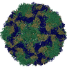
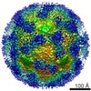
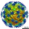
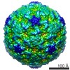
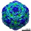
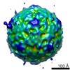
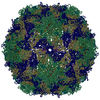
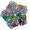
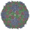
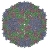
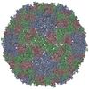
| 登録情報 | データベース: PDB / ID: 3j2j | ||||||
|---|---|---|---|---|---|---|---|
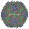
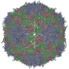
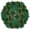
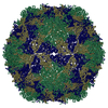
| タイトル | Empty coxsackievirus A9 capsid | ||||||
 要素 要素 |
| ||||||
 キーワード キーワード |  VIRUS (ウイルス) / CVA9-integrin / picornavirus (ピコルナウイルス科) / enterovirus (エンテロウイルス) / empty capsid VIRUS (ウイルス) / CVA9-integrin / picornavirus (ピコルナウイルス科) / enterovirus (エンテロウイルス) / empty capsid | ||||||
| 機能・相同性 |  機能・相同性情報 機能・相同性情報symbiont-mediated suppression of host cytoplasmic pattern recognition receptor signaling pathway via inhibition of RIG-I activity / ピコルナイン2A / symbiont-mediated suppression of host mRNA export from nucleus / symbiont genome entry into host cell via pore formation in plasma membrane / picornain 3C / T=pseudo3 icosahedral viral capsid / host cell cytoplasmic vesicle membrane / endocytosis involved in viral entry into host cell / : / nucleoside-triphosphate phosphatase ...symbiont-mediated suppression of host cytoplasmic pattern recognition receptor signaling pathway via inhibition of RIG-I activity / ピコルナイン2A / symbiont-mediated suppression of host mRNA export from nucleus / symbiont genome entry into host cell via pore formation in plasma membrane / picornain 3C / T=pseudo3 icosahedral viral capsid / host cell cytoplasmic vesicle membrane / endocytosis involved in viral entry into host cell / : / nucleoside-triphosphate phosphatase / protein complex oligomerization / monoatomic ion channel activity / RNA helicase activity / DNA複製 / induction by virus of host autophagy / RNA依存性RNAポリメラーゼ / symbiont-mediated suppression of host gene expression / viral RNA genome replication / cysteine-type endopeptidase activity / RNA-dependent RNA polymerase activity / DNA-templated transcription / host cell nucleus / virion attachment to host cell / structural molecule activity / ATP hydrolysis activity / タンパク質分解 / RNA binding / ATP binding / 生体膜 / metal ion binding類似検索 - 分子機能 | ||||||
| 生物種 |  Human coxsackievirus A9 (コクサッキーウイルス) Human coxsackievirus A9 (コクサッキーウイルス) | ||||||
| 手法 | 電子顕微鏡法 / 単粒子再構成法 / クライオ電子顕微鏡法 / 解像度: 9.54 Å | ||||||
 データ登録者 データ登録者 | Shakeel, S. / Seitsonen, J.J.T. / Kajander, T. / Laurinmaki, P. / Hyypia, T. / Susi, P. / Butcher, S.J. | ||||||
 引用 引用 | ジャーナル: J Virol / 年: 2013 タイトル: Structural and functional analysis of coxsackievirus A9 integrin αvβ6 binding and uncoating. 著者: Shabih Shakeel / Jani J T Seitsonen / Tommi Kajander / Pasi Laurinmäki / Timo Hyypiä / Petri Susi / Sarah J Butcher /  要旨: Coxsackievirus A9 (CVA9) is an important pathogen of the Picornaviridae family. It utilizes cellular receptors from the integrin αv family for binding to its host cells prior to entry and genome ...Coxsackievirus A9 (CVA9) is an important pathogen of the Picornaviridae family. It utilizes cellular receptors from the integrin αv family for binding to its host cells prior to entry and genome release. Among the integrins tested, it has the highest affinity for αvβ6, which recognizes the arginine-glycine-aspartic acid (RGD) loop present on the C terminus of viral capsid protein, VP1. As the atomic model of CVA9 lacks the RGD loop, we used surface plasmon resonance, electron cryo-microscopy, and image reconstruction to characterize the capsid-integrin interactions and the conformational changes on genome release. We show that the integrin binds to the capsid with nanomolar affinity and that the binding of integrin to the virion does not induce uncoating, thereby implying that further steps are required for release of the genome. Electron cryo-tomography and single-particle image reconstruction revealed variation in the number and conformation of the integrins bound to the capsid, with the integrin footprint mapping close to the predicted site for the exposed RGD loop on VP1. Comparison of empty and RNA-filled capsid reconstructions showed that the capsid undergoes conformational changes when the genome is released, so that the RNA-capsid interactions in the N termini of VP1 and VP4 are lost, VP4 is removed, and the capsid becomes more porous, as has been reported for poliovirus 1, human rhinovirus 2, enterovirus 71, and coxsackievirus A7. These results are important for understanding the structural basis of integrin binding to CVA9 and the molecular events leading to CVA9 cell entry and uncoating. | ||||||
| 履歴 |
|
- 構造の表示
構造の表示
| ムービー |
ムービービューア |
|---|---|
| 構造ビューア | 分子: MolmilJmol/JSmol |
- ダウンロードとリンク
-ダウンロード
| PDBx/mmCIF形式 | 3j2j.cif.gz | 134.9 KB | 表示 | PDBx/mmCIF形式 |
|---|---|---|---|---|
| PDB形式 | pdb3j2j.ent.gz | 100.8 KB | 表示 | PDB形式 |
| PDBx/mmJSON形式 | 3j2j.json.gz | ツリー表示 | PDBx/mmJSON形式 | |
| その他 |  その他のダウンロード その他のダウンロード |
-検証レポート
| アーカイブディレクトリ | https://data.pdbj.org/pub/pdb/validation_reports/j2/3j2jftp://data.pdbj.org/pub/pdb/validation_reports/j2/3j2j | HTTPS FTP |
|---|
-関連構造データ
-リンク
 PDBj
PDBj

- 集合体
集合体
| 登録構造単位 | 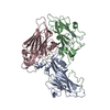
|
|---|---|
| 1 | x 60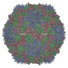
|
| 2 |
|
| 3 | x 5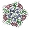
|
| 4 | x 6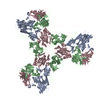
|
| 5 | 
|
| 対称性 | 点対称性: (シェーンフリース記号: I (正20面体型対称)) |
-要素
| #1: タンパク質 | 分子量: 25541.980 Da / 分子数: 1 / 断片: UNP residues 631-852 / 由来タイプ: 天然 由来: (天然) Human coxsackievirus A9 (コクサッキーウイルス)参照: UniProt: P21404 |
|---|---|
| #2: タンパク質 | 分子量: 26335.072 Da / 分子数: 1 / 断片: UNP residues 331-568 / 由来タイプ: 天然 由来: (天然) Human coxsackievirus A9 (コクサッキーウイルス)参照: UniProt: P21404 |
| #3: タンパク質 | 分子量: 27919.494 Da / 分子数: 1 / 断片: UNP residues 79-330 / 由来タイプ: 天然 由来: (天然) Human coxsackievirus A9 (コクサッキーウイルス)参照: UniProt: P21404 |
-実験情報
-実験
| 実験 | 手法: 電子顕微鏡法 |
|---|---|
| EM実験 | 試料の集合状態: PARTICLE / 3次元再構成法: 単粒子再構成法 |
- 試料調製
試料調製
| 構成要素 | 名称: Empty coxsackievirus A9 capsid in complex with integrin alpha v beta 6 タイプ: COMPLEX / 別称: CVA9 |
|---|---|
| ウイルスについての詳細 | 中空か: YES / エンベロープを持つか: NO / ホストのカテゴリ: VERTEBRATES / 単離: SPECIES / タイプ: VIRION |
| 天然宿主 | 生物種: Homo sapiens |
| 試料 | 包埋: NO / シャドウイング: NO / 染色: NO / 凍結: YES |
| 急速凍結 | 装置: GATAN CRYOPLUNGE 3 / 凍結剤: ETHANE 詳細: manual plunging into liquid ethane (GATAN CRYOPLUNGE 3) 手法: manual plunging |
- 電子顕微鏡撮影
| 実験機器 |  モデル: Tecnai F20 / 画像提供: FEI Company |
|---|---|
| 顕微鏡 | モデル: FEI TECNAI F20 / 日付: 2011年2月1日 |
| 電子銃 | 電子線源: FIELD EMISSION GUN / 加速電圧: 200 kV / 照射モード: FLOOD BEAM |
| 電子レンズ | モード: BRIGHT FIELDBright-field microscopy / 倍率(公称値): 62000 X / 倍率(補正後): 62000 X / 最大 デフォーカス(公称値): 4120 nm / 最小 デフォーカス(公称値): 830 nm / カメラ長: 0 mm |
| 試料ホルダ | 試料ホルダーモデル: GATAN LIQUID NITROGEN / 資料ホルダタイプ: GATAN 626 / 傾斜角・最大: 0 ° / 傾斜角・最小: 0 ° |
| 放射 | プロトコル: SINGLE WAVELENGTH / 単色(M)・ラウエ(L): M / 散乱光タイプ: x-ray |
| 放射波長 | 相対比: 1 |
- 解析
解析
| EMソフトウェア |
| ||||||||||||
|---|---|---|---|---|---|---|---|---|---|---|---|---|---|
| CTF補正 | 詳細: whole micrograph | ||||||||||||
| 対称性 | 点対称性: I (正20面体型対称) | ||||||||||||
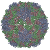
| 3次元再構成 | 手法: polar Fourier transform / 解像度: 9.54 Å / 解像度の算出法: FSC 0.5 CUT-OFF / 粒子像の数: 1200 / ピクセルサイズ(公称値): 2.26 Å / ピクセルサイズ(実測値): 2.26 Å / 対称性のタイプ: POINT | ||||||||||||
| 原子モデル構築 | プロトコル: FLEXIBLE FIT / 空間: REAL / 詳細: REFINEMENT PROTOCOL--flexible fitting | ||||||||||||
| 原子モデル構築 | 3D fitting-ID: 1 / Accession code: 1D4M / Initial refinement model-ID: 1 / PDB-ID: 1D4M / Source name: PDB / タイプ: experimental model
| ||||||||||||
| 精密化ステップ | サイクル: LAST
|