 ムービー
ムービー コントローラー


+ データを開く

- 基本情報
基本情報
| 登録情報 | データベース: EMDB / ID: EMD-6956 | |||||||||
|---|---|---|---|---|---|---|---|---|---|---|
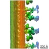
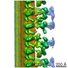
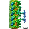
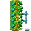
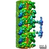
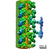
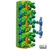
| タイトル | Doublet microtubule of zebrafish sperm axoneme, pih1d2_null mutant | |||||||||
 マップデータ マップデータ | doublet microtubule structure from zebrafish sperm flagella, pih1d2_null mutant | |||||||||
 試料 試料 |
| |||||||||
| 生物種 |   Danio rerio (ゼブラフィッシュ) Danio rerio (ゼブラフィッシュ) | |||||||||
| 手法 | 電子線トモグラフィー法 / クライオ電子顕微鏡法 / 解像度: 45.1 Å | |||||||||
 データ登録者 データ登録者 | Yamaguchi H / Oda T / Kikkawa M / Takeda H | |||||||||
 引用 引用 | ジャーナル: Elife / 年: 2018 タイトル: Systematic studies of all PIH proteins in zebrafish reveal their distinct roles in axonemal dynein assembly. 著者: Hiroshi Yamaguchi / Toshiyuki Oda / Masahide Kikkawa / Hiroyuki Takeda /  要旨: Construction of motile cilia/flagella requires cytoplasmic preassembly of axonemal dyneins before transport into cilia. Axonemal dyneins have various subtypes, but the roles of each dynein subtype ...Construction of motile cilia/flagella requires cytoplasmic preassembly of axonemal dyneins before transport into cilia. Axonemal dyneins have various subtypes, but the roles of each dynein subtype and their assembly processes remain elusive in vertebrates. The PIH protein family, consisting of four members, has been implicated in the assembly of different dynein subtypes, although evidence for this idea is sparse. Here, we established zebrafish mutants of all four PIH-protein genes: , , , and , and analyzed the structures of axonemal dyneins in mutant spermatozoa by cryo-electron tomography. Mutations caused the loss of specific dynein subtypes, which was correlated with abnormal sperm motility. We also found organ-specific compositions of dynein subtypes, which could explain the severe motility defects of mutant Kupffer's vesicle cilia. Our data demonstrate that all vertebrate PIH proteins are differently required for cilia/flagella motions and the assembly of axonemal dyneins, assigning specific dynein subtypes to each PIH protein. | |||||||||
| 履歴 |
|
- 構造の表示
構造の表示
| ムービー |
 ムービービューア ムービービューア |
|---|---|
| 構造ビューア | EMマップ: SurfViewMolmilJmol/JSmol |
| 添付画像 |

- ダウンロードとリンク
-EMDBアーカイブ
| マップデータ | emd_6956.map.gz | 9.1 MB | EMDBマップデータ形式 | |
|---|---|---|---|---|
| ヘッダ (付随情報) | emd-6956-v30.xmlemd-6956.xml | 10.6 KB 10.6 KB | 表示 表示 | EMDBヘッダ |
| FSC (解像度算出) | emd_6956_fsc.xml | 6.4 KB | 表示 | FSCデータファイル |
| 画像 | 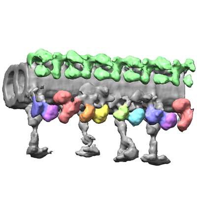 emd_6956.png emd_6956.png | 106.9 KB | ||
| アーカイブディレクトリ |  http://ftp.pdbj.org/pub/emdb/structures/EMD-6956ftp://ftp.pdbj.org/pub/emdb/structures/EMD-6956 http://ftp.pdbj.org/pub/emdb/structures/EMD-6956ftp://ftp.pdbj.org/pub/emdb/structures/EMD-6956 | HTTPS FTP |
-関連構造データ

-リンク
| EMDBのページ | EMDB (EBI/PDBe) / EMDataResource |
|---|
-マップ
| ファイル | ダウンロード / ファイル: emd_6956.map.gz / 形式: CCP4 / 大きさ: 9.9 MB / タイプ: IMAGE STORED AS FLOATING POINT NUMBER (4 BYTES) | ||||||||||||||||||||||||||||||||||||||||||||||||||||||||||||||||||||
|---|---|---|---|---|---|---|---|---|---|---|---|---|---|---|---|---|---|---|---|---|---|---|---|---|---|---|---|---|---|---|---|---|---|---|---|---|---|---|---|---|---|---|---|---|---|---|---|---|---|---|---|---|---|---|---|---|---|---|---|---|---|---|---|---|---|---|---|---|---|
| 注釈 | doublet microtubule structure from zebrafish sperm flagella, pih1d2_null mutant | ||||||||||||||||||||||||||||||||||||||||||||||||||||||||||||||||||||
| ボクセルのサイズ | X=Y=Z: 7.2 Å | ||||||||||||||||||||||||||||||||||||||||||||||||||||||||||||||||||||
| 密度 |
| ||||||||||||||||||||||||||||||||||||||||||||||||||||||||||||||||||||
| 対称性 | 空間群: 1 | ||||||||||||||||||||||||||||||||||||||||||||||||||||||||||||||||||||
| 詳細 | EMDB XML:
CCP4マップ ヘッダ情報:
| ||||||||||||||||||||||||||||||||||||||||||||||||||||||||||||||||||||
-添付データ
- 試料の構成要素
-全体 : Doublet microtubule from zebrafish sperm flagella, pih1d2_null mutant
| 全体 | 名称: Doublet microtubule from zebrafish sperm flagella, pih1d2_null mutant |
|---|---|
| 要素 |
|
-超分子 #1: Doublet microtubule from zebrafish sperm flagella, pih1d2_null mutant
| 超分子 | 名称: Doublet microtubule from zebrafish sperm flagella, pih1d2_null mutant タイプ: organelle_or_cellular_component / ID: 1 / 親要素: 0 |
|---|---|
| 由来(天然) | 生物種: Danio rerio (ゼブラフィッシュ) / Organelle: flagella |
-実験情報
-構造解析
| 手法 | クライオ電子顕微鏡法 |
|---|---|
 解析 解析 | 電子線トモグラフィー法 |
| 試料の集合状態 | filament |
-試料調製
| 緩衝液 | pH: 7.2 | ||||||
|---|---|---|---|---|---|---|---|
| グリッド | モデル: Homemade / 材質: COPPER / メッシュ: 300 / 支持フィルム - 材質: CARBON / 支持フィルム - トポロジー: HOLEY / 前処理 - タイプ: GLOW DISCHARGE | ||||||
| 凍結 | 凍結剤: ETHANE / 装置: LEICA EM GP | ||||||
| 切片作成 | その他: NO SECTIONING | ||||||
| 位置合わせマーカー |
|
- 電子顕微鏡法
| 顕微鏡 | JEOL 3100FFC |
|---|---|
| 電子線 | 加速電圧: 300 kV / 電子線源: FIELD EMISSION GUN |
| 電子光学系 | 照射モード: FLOOD BEAM / 撮影モード: BRIGHT FIELDBright-field microscopy / 倍率(公称値): 30000 |
| 特殊光学系 | エネルギーフィルター - 名称: In-column Omega Filter |
| 試料ステージ | 試料ホルダーモデル: GATAN 914 HIGH TILT LIQUID NITROGEN CRYO TRANSFER TOMOGRAPHY HOLDER ホルダー冷却材: NITROGEN |
| 撮影 | フィルム・検出器のモデル: TVIPS TEMCAM-F416 (4k x 4k) デジタル化 - サイズ - 横: 4096 pixel / デジタル化 - サイズ - 縦: 4096 pixel / 平均電子線量: 1.6 e/Å2 |
-画像解析
| CTF補正 | ソフトウェア - 名称: IMOD |
|---|---|
| 最終 再構成 | アルゴリズム: BACK PROJECTION / 解像度のタイプ: BY AUTHOR / 解像度: 45.1 Å / 解像度の算出法: FSC 0.143 CUT-OFF / ソフトウェア - 名称: IMOD / 使用した粒子像数: 60 |
| FSC曲線 (解像度の算出) |  |
