Mitochondrial mRNA-processing protein COX24, C-terminal / Mitochondrial mRNA-processing protein COX24, C-terminal / Mitochondrial domain of unknown function (DUF1713) / Ribosomal protein S14, type Z / Ribosomal protein S16, conserved site / Ribosomal protein S16 signature. / Ribosomal protein S14/S29 / Ribosomal protein S3, bacterial-type / Ribosomal protein S6, conserved site / Ribosomal protein S6 signature. ...Mitochondrial mRNA-processing protein COX24, C-terminal / Mitochondrial mRNA-processing protein COX24, C-terminal / Mitochondrial domain of unknown function (DUF1713) / Ribosomal protein S14, type Z / Ribosomal protein S16, conserved site / Ribosomal protein S16 signature. / Ribosomal protein S14/S29 / Ribosomal protein S3, bacterial-type / Ribosomal protein S6, conserved site / Ribosomal protein S6 signature. / Ribosomal protein S19, bacterial-type / Ribosomal protein S7, bacterial/organellar-type / Ribosomal protein S11, bacterial-type / Ribosomal protein S13, bacterial-type / Ribosomal protein S20 / Ribosomal protein S20 superfamily / Ribosomal protein S20 / Ribosomal protein S9, bacterial/plastid / Ribosomal protein S4, bacterial-type / 30S ribosomal protein S17 / Ribosomal protein S5, bacterial-type / Ribosomal protein S6, plastid/chloroplast / Ribosomal protein S2, bacteria/mitochondria/plastid / Ribosomal protein S18, conserved site / Ribosomal protein S18 signature. / Ribosomal protein S16 / Ribosomal protein S16 / Ribosomal protein S16 domain superfamily / Ribosomal protein S15, bacterial-type / Ribosomal protein S6 / Ribosomal protein S6 / Ribosomal protein S6 superfamily / Ribosomal protein S12, bacterial-type / Translation elongation factor EF1B/ribosomal protein S6 / Ribosomal protein S18 / Ribosomal protein S18 / Ribosomal protein S18 superfamily / KHドメイン / K homology RNA-binding domain / Ribosomal protein S3, conserved site / Ribosomal protein S14, conserved site / Ribosomal protein S10, conserved site / : / K Homology domain, type 2 / Ribosomal protein S3, C-terminal / Ribosomal protein S3, C-terminal domain superfamily / Ribosomal protein S15/S19, conserved site / KHドメイン / Ribosomal protein S19/S15 / Ribosomal protein S19/S15, superfamily / Ribosomal protein S10 / Ribosomal protein S3, C-terminal domain / Ribosomal protein S3 signature. / Ribosomal protein S10 signature. / Ribosomal protein S14 signature. / Ribosomal protein S7, conserved site / Ribosomal protein S17, conserved site / K homology domain superfamily, prokaryotic type / Ribosomal protein S19 / Ribosomal protein S2 signature 1. / Ribosomal protein S13, conserved site / Ribosomal protein S2, conserved site / Ribosomal protein S13 / 30s ribosomal protein S13, C-terminal / Ribosomal protein S2 / Ribosomal protein S2, flavodoxin-like domain superfamily / Ribosomal protein S14 / Ribosomal protein S2 / Ribosomal protein S4/S9 N-terminal domain / Type-2 KH domain profile. / Ribosomal protein S4/S9, N-terminal / Ribosomal protein S4, conserved site / Ribosomal protein S4/S9 N-terminal domain / Ribosomal protein S13/S18 / Ribosomal protein S4/S9 / Ribosomal protein S19 signature. / K homology domain-like, alpha/beta / Ribosomal protein S14p/S29e / Ribosomal protein S8 / Ribosomal protein S8 superfamily / Ribosomal protein S5, N-terminal, conserved site / Ribosomal protein S5 signature. / Ribosomal protein S7 signature. / Ribosomal protein S5 / Ribosomal protein S5, N-terminal / Ribosomal S11, conserved site / Ribosomal protein S10p/S20e / Ribosomal protein S13-like, H2TH / S5 double stranded RNA-binding domain profile. / Ribosomal protein S5, C-terminal / Ribosomal protein S9, conserved site / Ribosomal protein S5, N-terminal domain / Ribosomal protein S8 / Ribosomal protein S10 domain / Ribosomal protein S10 domain superfamily / Ribosomal protein S17 signature. / Ribosomal protein S5, C-terminal domain / S4 RNA-binding domain / Ribosomal protein S11 / RNA-binding S4 domain 類似検索 - ドメイン・相同性
30S ribosomal protein S6 / Conserved domain protein / Small ribosomal subunit protein uS12 / Small ribosomal subunit protein uS7 / Small ribosomal subunit protein uS10 / Small ribosomal subunit protein uS19 / Small ribosomal subunit protein uS3 / Small ribosomal subunit protein uS17 / Small ribosomal subunit protein uS14B / Small ribosomal subunit protein uS8 ...30S ribosomal protein S6 / Conserved domain protein / Small ribosomal subunit protein uS12 / Small ribosomal subunit protein uS7 / Small ribosomal subunit protein uS10 / Small ribosomal subunit protein uS19 / Small ribosomal subunit protein uS3 / Small ribosomal subunit protein uS17 / Small ribosomal subunit protein uS14B / Small ribosomal subunit protein uS8 / Small ribosomal subunit protein uS5 / Small ribosomal subunit protein uS13 / Small ribosomal subunit protein uS11 / Small ribosomal subunit protein uS4 / Small ribosomal subunit protein uS9 / Small ribosomal subunit protein bS16 / Small ribosomal subunit protein uS2 / Small ribosomal subunit protein uS15 / Small ribosomal subunit protein bS20 / Small ribosomal subunit protein bS18B 類似検索 - 構成要素
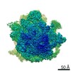
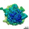
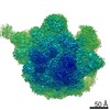
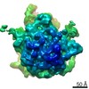
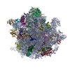
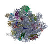
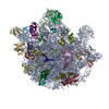
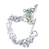
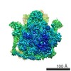
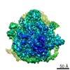
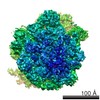
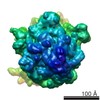
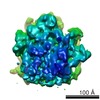
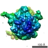
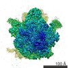
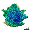
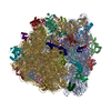
ジャーナル: Sci Rep / 年: 2018 タイトル: Structures of Mycobacterium smegmatis 70S ribosomes in complex with HPF, tmRNA, and P-tRNA. 著者: Satabdi Mishra / Tofayel Ahmed / Anu Tyagi / Jian Shi / Shashi Bhushan / 要旨: Ribosomes are the dynamic protein synthesis machineries of the cell. They may exist in different functional states in the cell. Therefore, it is essential to have structural information on these ...Ribosomes are the dynamic protein synthesis machineries of the cell. They may exist in different functional states in the cell. Therefore, it is essential to have structural information on these different functional states of ribosomes to understand their mechanism of action. Here, we present single particle cryo-EM reconstructions of the Mycobacterium smegmatis 70S ribosomes in the hibernating state (with HPF), trans-translating state (with tmRNA), and the P/P state (with P-tRNA) resolved to 4.1, 12.5, and 3.4 Å, respectively. A comparison of the P/P state with the hibernating state provides possible functional insights about the Mycobacteria-specific helix H54a rRNA segment. Interestingly, densities for all the four OB domains of bS1 protein is visible in the hibernating 70S ribosome displaying the molecular details of bS1-70S interactions. Our structural data shows a Mycobacteria-specific H54a-bS1 interaction which seems to prevent subunit dissociation and degradation during hibernation without the formation of 100S dimer. This indicates a new role of bS1 protein in 70S protection during hibernation in Mycobacteria in addition to its conserved function during translation initiation.
 ムービー
ムービー コントローラー


 データを開く
 基本情報
基本情報 マップデータ
 試料
試料 機能・相同性情報
 tRNA binding /
  Mycobacterium smegmatis str. MC2 155 (バクテリア) /
  データ登録者
 シンガポール, 1件
  引用
引用 構造の表示
構造の表示
 ダウンロードとリンク
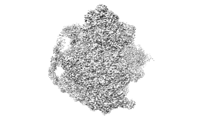 emd_6923.png
 http://ftp.pdbj.org/pub/emdb/structures/EMD-6923
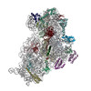


 試料の構成要素
 解析
解析 電子顕微鏡法
