 ムービー
ムービー コントローラー


+ データを開く

- 基本情報
基本情報
| 登録情報 | データベース: EMDB / ID: EMD-5730 | |||||||||
|---|---|---|---|---|---|---|---|---|---|---|
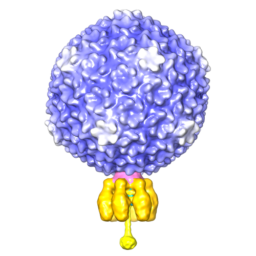
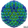
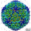
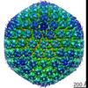
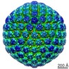
| タイトル | Asymmetric reconstruction of phage Sf6 | |||||||||
 マップデータ マップデータ | asymmetric reconstruction of phage Sf6 | |||||||||
 試料 試料 |
| |||||||||
| 生物種 |  Shigella phage Sf6 (ファージ) Shigella phage Sf6 (ファージ) | |||||||||
| 手法 |  単粒子再構成法 / クライオ電子顕微鏡法 / 解像度: 16.0 Å 単粒子再構成法 / クライオ電子顕微鏡法 / 解像度: 16.0 Å | |||||||||
 データ登録者 データ登録者 | Parent KN / Gilcrease EB / Casjens SR / Baker TS | |||||||||
 引用 引用 | ジャーナル: Virology / 年: 2012 タイトル: Structural evolution of the P22-like phages: comparison of Sf6 and P22 procapsid and virion architectures. 著者: Kristin N Parent / Eddie B Gilcrease / Sherwood R Casjens / Timothy S Baker /  要旨: Coat proteins of tailed, dsDNA phages and in herpesviruses include a conserved core similar to the bacteriophage HK97 subunit. This core is often embellished with other domains such as the telokin Ig- ...Coat proteins of tailed, dsDNA phages and in herpesviruses include a conserved core similar to the bacteriophage HK97 subunit. This core is often embellished with other domains such as the telokin Ig-like domain of phage P22. Eighty-six P22-like phages and prophages with sequenced genomes share a similar set of virion assembly genes and, based on comparisons of twelve viral assembly proteins (structural and assembly/packaging chaperones), these phages are classified into three groups (P22-like, Sf6-like, and CUS-3-like). We used cryo-electron microscopy and 3D image reconstruction to determine the structures of Sf6 procapsids and virions (~7Å resolution), and the structure of the entire, asymmetric Sf6 virion (16-Å resolution). The Sf6 coat protein is similar to that of P22 yet it has differences in the telokin domain and in its overall quaternary organization. Thermal stability and agarose gel experiments show that Sf6 virions are slightly less stable than those of P22. Finally, bacterial host outer membrane proteins A and C were identified in lipid vesicles that co-purify with Sf6 particles, but are not components of the capsid. | |||||||||
| 履歴 |
|
- 構造の表示
構造の表示
| ムービー |
 ムービービューア ムービービューア |
|---|---|
| 構造ビューア | EMマップ: SurfViewMolmilJmol/JSmol |
| 添付画像 |
- ダウンロードとリンク
-EMDBアーカイブ
| マップデータ | emd_5730.map.gz | 446 MB | EMDBマップデータ形式 | |
|---|---|---|---|---|
| ヘッダ (付随情報) | emd-5730-v30.xmlemd-5730.xml | 9.3 KB 9.3 KB | 表示 表示 | EMDBヘッダ |
| 画像 |  emd_5730_1.png emd_5730_1.png | 173.8 KB | ||
| アーカイブディレクトリ |  http://ftp.pdbj.org/pub/emdb/structures/EMD-5730ftp://ftp.pdbj.org/pub/emdb/structures/EMD-5730 http://ftp.pdbj.org/pub/emdb/structures/EMD-5730ftp://ftp.pdbj.org/pub/emdb/structures/EMD-5730 | HTTPS FTP |
-関連構造データ
-リンク
| EMDBのページ | EMDB (EBI/PDBe) / EMDataResource |
|---|
-マップ
| ファイル | ダウンロード / ファイル: emd_5730.map.gz / 形式: CCP4 / 大きさ: 1.3 GB / タイプ: IMAGE STORED AS FLOATING POINT NUMBER (4 BYTES) | ||||||||||||||||||||||||||||||||||||||||||||||||||||||||||||||||||||
|---|---|---|---|---|---|---|---|---|---|---|---|---|---|---|---|---|---|---|---|---|---|---|---|---|---|---|---|---|---|---|---|---|---|---|---|---|---|---|---|---|---|---|---|---|---|---|---|---|---|---|---|---|---|---|---|---|---|---|---|---|---|---|---|---|---|---|---|---|---|
| 注釈 | asymmetric reconstruction of phage Sf6 | ||||||||||||||||||||||||||||||||||||||||||||||||||||||||||||||||||||
| ボクセルのサイズ | X=Y=Z: 2.14 Å | ||||||||||||||||||||||||||||||||||||||||||||||||||||||||||||||||||||
| 密度 |
| ||||||||||||||||||||||||||||||||||||||||||||||||||||||||||||||||||||
| 対称性 | 空間群: 1 | ||||||||||||||||||||||||||||||||||||||||||||||||||||||||||||||||||||
| 詳細 | EMDB XML:
CCP4マップ ヘッダ情報:
| ||||||||||||||||||||||||||||||||||||||||||||||||||||||||||||||||||||
-添付データ
- 試料の構成要素
-全体 : phage sf6 mature virion
| 全体 | 名称: phage sf6 mature virion |
|---|---|
| 要素 |
|
-超分子 #1000: phage sf6 mature virion
| 超分子 | 名称: phage sf6 mature virion / タイプ: sample / ID: 1000 集合状態: icosahedral head, trimeric tail needle, 6-fold tailspikes, 12-fold portal and gp4 Number unique components: 14 |
|---|
-超分子 #1: Shigella phage Sf6
| 超分子 | 名称: Shigella phage Sf6 / タイプ: virus / ID: 1 / NCBI-ID: 10761 / 生物種: Shigella phage Sf6 / Sci species strain: clear mutant / データベース: NCBI / ウイルスタイプ: VIRION / ウイルス・単離状態: STRAIN / ウイルス・エンベロープ: No / ウイルス・中空状態: No |
|---|---|
| 宿主 | 生物種:  Shigella flexneri (フレクスナー赤痢菌) / 株: PE577 / 別称: BACTERIA(EUBACTERIA) Shigella flexneri (フレクスナー赤痢菌) / 株: PE577 / 別称: BACTERIA(EUBACTERIA) |
-実験情報
-構造解析
| 手法 | クライオ電子顕微鏡法 |
|---|---|
 解析 解析 | 単粒子再構成法 |
| 試料の集合状態 | particle |
-試料調製
| 濃度 | 10 mg/mL |
|---|---|
| 緩衝液 | pH: 7.6 / 詳細: 10mM Tris, 10mM MgCl2 |
| グリッド | 詳細: 400 mesh Quantifoil R2/2 |
| 凍結 | 凍結剤: ETHANE / チャンバー内湿度: 99 % / チャンバー内温度: 90 K / 装置: HOMEMADE PLUNGER / 手法: Blot for 5 sec before plunging. |
- 電子顕微鏡法
| 顕微鏡 | FEI POLARA 300 |
|---|---|
| 電子線 | 加速電圧: 200 kV / 電子線源: FIELD EMISSION GUN |
| 電子光学系 | 倍率(補正後): 58050 / 照射モード: FLOOD BEAM / 撮影モード: BRIGHT FIELDBright-field microscopy / Cs: 2.3 mm / 最大 デフォーカス(公称値): 4.61 µm / 最小 デフォーカス(公称値): 0.1 µm / 倍率(公称値): 59000 |
| 試料ステージ | 試料ホルダーモデル: OTHER |
| 温度 | 最低: 89 K / 最高: 91 K / 平均: 90 K |
| アライメント法 | Legacy - 非点収差: objective lens astigmatism was corrected at high magnification |
| 日付 | 2010年9月30日 |
| 撮影 | カテゴリ: FILM / フィルム・検出器のモデル: KODAK SO-163 FILM / デジタル化 - スキャナー: OTHER / デジタル化 - サンプリング間隔: 1.07 µm / 実像数: 611 / 平均電子線量: 22 e/Å2 / ビット/ピクセル: 8 |
| 実験機器 |  モデル: Tecnai Polara / 画像提供: FEI Company |
-画像解析
| CTF補正 | 詳細: Robem |
|---|---|
| 最終 再構成 | アルゴリズム: OTHER / 解像度のタイプ: BY AUTHOR / 解像度: 16.0 Å / 解像度の算出法: FSC 0.5 CUT-OFF / ソフトウェア - 名称: Auto3dem / 使用した粒子像数: 16253 |
| 詳細 | the reconstruction was done using Auto3dem. |
