 ムービー
ムービー コントローラー


+ データを開く

- 基本情報
基本情報
| 登録情報 | データベース: EMDB / ID: EMD-5689 | |||||||||
|---|---|---|---|---|---|---|---|---|---|---|
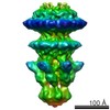
| タイトル | Cryo-electron microscopy of T7 tail complex | |||||||||
 マップデータ マップデータ | Reconstruction of T7 tail complex | |||||||||
 試料 試料 |
| |||||||||
 キーワード キーワード |  Bacteriophage (ファージ) / DNA ejection / tail complex (尾) / connector / gatekeeper Bacteriophage (ファージ) / DNA ejection / tail complex (尾) / connector / gatekeeper | |||||||||
| 生物種 |   Enterobacteria phage T7 (ファージ) Enterobacteria phage T7 (ファージ) | |||||||||
| 手法 | 単粒子再構成法 / クライオ電子顕微鏡法 / 解像度: 16.0 Å | |||||||||
 データ登録者 データ登録者 | Cuervo A / Pulido-Cid M / Chagoyen M / Arranz R / Gonzalez-Garcia VA / Garcia-Doval C / Caston JR / Valpuesta JM / van Raaij MJ / Martin-Benito J / Carrascosa JL | |||||||||
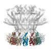
 引用 引用 | ジャーナル: J Biol Chem / 年: 2013 タイトル: Structural characterization of the bacteriophage T7 tail machinery. 著者: Ana Cuervo / Mar Pulido-Cid / Mónica Chagoyen / Rocío Arranz / Verónica A González-García / Carmela Garcia-Doval / José R Castón / José M Valpuesta / Mark J van Raaij / Jaime Martín- ...著者: Ana Cuervo / Mar Pulido-Cid / Mónica Chagoyen / Rocío Arranz / Verónica A González-García / Carmela Garcia-Doval / José R Castón / José M Valpuesta / Mark J van Raaij / Jaime Martín-Benito / José L Carrascosa /  要旨: Most bacterial viruses need a specialized machinery, called "tail," to inject their genomes inside the bacterial cytoplasm without disrupting the cellular integrity. Bacteriophage T7 is a well ...Most bacterial viruses need a specialized machinery, called "tail," to inject their genomes inside the bacterial cytoplasm without disrupting the cellular integrity. Bacteriophage T7 is a well characterized member of the Podoviridae family infecting Escherichia coli, and it has a short noncontractile tail that assembles sequentially on the viral head after DNA packaging. The T7 tail is a complex of around 2.7 MDa composed of at least four proteins as follows: the connector (gene product 8, gp8), the tail tubular proteins gp11 and gp12, and the fibers (gp17). Using cryo-electron microscopy and single particle image reconstruction techniques, we have determined the precise topology of the tail proteins by comparing the structure of the T7 tail extracted from viruses and a complex formed by recombinant gp8, gp11, and gp12 proteins. Furthermore, the order of assembly of the structural components within the complex was deduced from interaction assays with cloned and purified tail proteins. The existence of common folds among similar tail proteins allowed us to obtain pseudo-atomic threaded models of gp8 (connector) and gp11 (gatekeeper) proteins, which were docked into the corresponding cryo-EM volumes of the tail complex. This pseudo-atomic model of the connector-gatekeeper interaction revealed the existence of a common molecular architecture among viruses belonging to the three tailed bacteriophage families, strongly suggesting that a common molecular mechanism has been favored during evolution to coordinate the transition between DNA packaging and tail assembly. | |||||||||
| 履歴 |
|
- 構造の表示
構造の表示
| ムービー |
 ムービービューア ムービービューア |
|---|---|
| 構造ビューア | EMマップ: SurfViewMolmilJmol/JSmol |
| 添付画像 |

- ダウンロードとリンク
-EMDBアーカイブ
| マップデータ | emd_5689.map.gz | 14.2 MB | EMDBマップデータ形式 | |
|---|---|---|---|---|
| ヘッダ (付随情報) | emd-5689-v30.xmlemd-5689.xml | 12.2 KB 12.2 KB | 表示 表示 | EMDBヘッダ |
| 画像 | 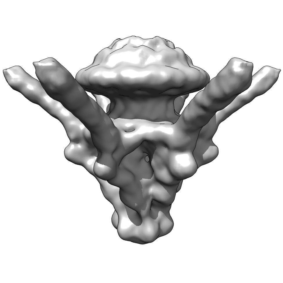 emd_5689_1.jpg emd_5689_1.jpg | 76.3 KB | ||
| アーカイブディレクトリ |  http://ftp.pdbj.org/pub/emdb/structures/EMD-5689ftp://ftp.pdbj.org/pub/emdb/structures/EMD-5689 http://ftp.pdbj.org/pub/emdb/structures/EMD-5689ftp://ftp.pdbj.org/pub/emdb/structures/EMD-5689 | HTTPS FTP |
-関連構造データ

-リンク
| EMDBのページ | EMDB (EBI/PDBe) / EMDataResource |
|---|
-マップ
| ファイル | ダウンロード / ファイル: emd_5689.map.gz / 形式: CCP4 / 大きさ: 15.3 MB / タイプ: IMAGE STORED AS FLOATING POINT NUMBER (4 BYTES) | ||||||||||||||||||||||||||||||||||||||||||||||||||||||||||||||||||||
|---|---|---|---|---|---|---|---|---|---|---|---|---|---|---|---|---|---|---|---|---|---|---|---|---|---|---|---|---|---|---|---|---|---|---|---|---|---|---|---|---|---|---|---|---|---|---|---|---|---|---|---|---|---|---|---|---|---|---|---|---|---|---|---|---|---|---|---|---|---|
| 注釈 | Reconstruction of T7 tail complex | ||||||||||||||||||||||||||||||||||||||||||||||||||||||||||||||||||||
| ボクセルのサイズ | X=Y=Z: 2.8 Å | ||||||||||||||||||||||||||||||||||||||||||||||||||||||||||||||||||||
| 密度 |
| ||||||||||||||||||||||||||||||||||||||||||||||||||||||||||||||||||||
| 対称性 | 空間群: 1 | ||||||||||||||||||||||||||||||||||||||||||||||||||||||||||||||||||||
| 詳細 | EMDB XML:
CCP4マップ ヘッダ情報:
| ||||||||||||||||||||||||||||||||||||||||||||||||||||||||||||||||||||
-添付データ
- 試料の構成要素
-全体 : T7 tail complex formed by proteins gp8, gp11, gp12, and gp17
| 全体 | 名称: T7 tail complex formed by proteins gp8, gp11, gp12, and gp17 |
|---|---|
| 要素 |
|
-超分子 #1000: T7 tail complex formed by proteins gp8, gp11, gp12, and gp17
| 超分子 | 名称: T7 tail complex formed by proteins gp8, gp11, gp12, and gp17 タイプ: sample / ID: 1000 集合状態: gp8 (12mer), gp11 (12mer), gp12 (6mer), gp17 (6mer of 3mers) Number unique components: 4 |
|---|---|
| 分子量 | 理論値: 2.7 MDa |
-分子 #1: gp8
| 分子 | 名称: gp8 / タイプ: protein_or_peptide / ID: 1 / Name.synonym: Connector / コピー数: 12 / 集合状態: 12mer / 組換発現: No / データベース: NCBI |
|---|---|
| 由来(天然) | 生物種: Enterobacteria phage T7 (ファージ) / 別称: T7 Bacteriophage |
| 分子量 | 理論値: 59 KDa |
-分子 #2: gp17
| 分子 | 名称: gp17 / タイプ: protein_or_peptide / ID: 2 / Name.synonym: Fibre / コピー数: 18 / 集合状態: 3mer / 組換発現: No / データベース: NCBI |
|---|---|
| 由来(天然) | 生物種: Enterobacteria phage T7 (ファージ) / 別称: T7 Bacteriophage |
| 分子量 | 理論値: 60 KDa |
-分子 #3: gp11
| 分子 | 名称: gp11 / タイプ: protein_or_peptide / ID: 3 / Name.synonym: gatekeeper / コピー数: 12 / 集合状態: 12mer / 組換発現: No / データベース: NCBI |
|---|---|
| 由来(天然) | 生物種: Enterobacteria phage T7 (ファージ) / 別称: T7 bacteriophage |
| 分子量 | 理論値: 25 KDa |
-分子 #4: gp12
| 分子 | 名称: gp12 / タイプ: protein_or_peptide / ID: 4 / コピー数: 6 / 集合状態: 6mer / 組換発現: No / データベース: NCBI |
|---|---|
| 由来(天然) | 生物種: Enterobacteria phage T7 (ファージ) / 別称: T7 bacteriophage |
| 分子量 | 理論値: 90 KDa |
-実験情報
-構造解析
| 手法 | クライオ電子顕微鏡法 |
|---|---|
 解析 解析 | 単粒子再構成法 |
| 試料の集合状態 | particle |
-試料調製
| 濃度 | 0.1 mg/mL |
|---|---|
| 緩衝液 | pH: 7.8 / 詳細: 50mM Tris-HCl, 10mM MgCl2, 100 mM NaCl |
| グリッド | 詳細: R2/2 Quantifoil coated with a thin carbon layer |
| 凍結 | 凍結剤: ETHANE / 装置: LEICA EM CPC 手法: Samples were applied to grids for 1 minute, blotted, and plunged into liquid ethane. |
- 電子顕微鏡法
| 顕微鏡 | FEI TECNAI F20 |
|---|---|
| 電子線 | 加速電圧: 200 kV / 電子線源: FIELD EMISSION GUN |
| 電子光学系 | 倍率(補正後): 50000 / 照射モード: FLOOD BEAM / 撮影モード: BRIGHT FIELDBright-field microscopy / Cs: 2.26 mm / 最大 デフォーカス(公称値): 3.5 µm / 最小 デフォーカス(公称値): 1.5 µm / 倍率(公称値): 50000 |
| 試料ステージ | 試料ホルダーモデル: GATAN LIQUID NITROGEN |
| 温度 | 最低: 91 K / 最高: 113 K |
| 日付 | 2009年2月1日 |
| 撮影 | カテゴリ: FILM / フィルム・検出器のモデル: KODAK SO-163 FILM / デジタル化 - スキャナー: ZEISS SCAI / デジタル化 - サンプリング間隔: 7 µm / 実像数: 22 / 平均電子線量: 10 e/Å2 / ビット/ピクセル: 8 |
| 実験機器 |  モデル: Tecnai F20 / 画像提供: FEI Company |
-画像解析
| CTF補正 | 詳細: each micrograph |
|---|---|
| 最終 再構成 | アルゴリズム: OTHER / 解像度のタイプ: BY AUTHOR / 解像度: 16.0 Å / 解像度の算出法: FSC 0.33 CUT-OFF / ソフトウェア - 名称: XMIPP, SPIDER, EMAN 詳細: The resolution is 20 Angstrom with FSC at 0.5 cut-off. 使用した粒子像数: 3056 |
