ムービー
ムービー コントローラー


+ データを開く

- 基本情報
基本情報
| 登録情報 | データベース: EMDB / ID: EMD-5405 | |||||||||
|---|---|---|---|---|---|---|---|---|---|---|
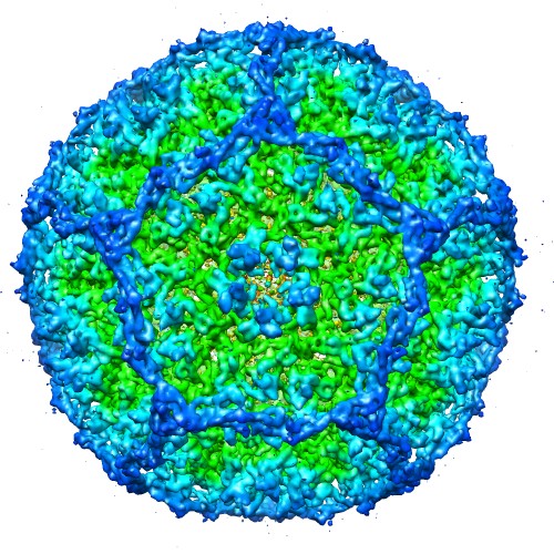
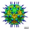
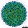
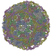
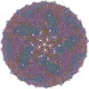
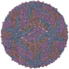
| タイトル | Icosahedral reconstruction of bacteriophage P4 procapsid | |||||||||
 マップデータ マップデータ | Reconstruction of P4 procapsid | |||||||||
 試料 試料 |
| |||||||||
 キーワード キーワード |  bacteriophage (ファージ) / P2 / P4 / capsid (カプシド) / procapsid (カプシド) / assembly / size determination / scaffolding protein (足場タンパク質) / triangulation numbers / T=7 dextro / Sid / gpN bacteriophage (ファージ) / P2 / P4 / capsid (カプシド) / procapsid (カプシド) / assembly / size determination / scaffolding protein (足場タンパク質) / triangulation numbers / T=7 dextro / Sid / gpN | |||||||||
| 生物種 |  Escherichia coli (大腸菌) Escherichia coli (大腸菌) | |||||||||
| 手法 | 単粒子再構成法 / クライオ電子顕微鏡法 / 解像度: 8.0 Å | |||||||||
 データ登録者 データ登録者 | Dearborn AD / Laurinmaki P / Chandramouli P / Rodenburg CM / Wang S / Butcher SJ / Dokland T | |||||||||
 引用 引用 | ジャーナル: J Struct Biol / 年: 2012 タイトル: Structure and size determination of bacteriophage P2 and P4 procapsids: function of size responsiveness mutations. 著者: Altaira D Dearborn / Pasi Laurinmaki / Preethi Chandramouli / Cynthia M Rodenburg / Sifang Wang / Sarah J Butcher / Terje Dokland /  要旨: Bacteriophage P4 is dependent on structural proteins supplied by a helper phage, P2, to assemble infectious virions. Bacteriophage P2 normally forms an icosahedral capsid with T=7 symmetry from the ...Bacteriophage P4 is dependent on structural proteins supplied by a helper phage, P2, to assemble infectious virions. Bacteriophage P2 normally forms an icosahedral capsid with T=7 symmetry from the gpN capsid protein, the gpO scaffolding protein and the gpQ portal protein. In the presence of P4, however, the same structural proteins are assembled into a smaller capsid with T=4 symmetry. This size determination is effected by the P4-encoded protein Sid, which forms an external scaffold around the small P4 procapsids. Size responsiveness (sir) mutants in gpN fail to assemble small capsids even in the presence of Sid. We have produced large and small procapsids by co-expression of gpN with gpO and Sid, respectively, and applied cryo-electron microscopy and three-dimensional reconstruction methods to visualize these procapsids. gpN has an HK97-like fold and interacts with Sid in an exposed loop where the sir mutations are clustered. The T=7 lattice of P2 has dextro handedness, unlike the laevo lattices of other phages with this fold observed so far. | |||||||||
| 履歴 |
|
- 構造の表示
構造の表示
| ムービー |
 ムービービューア ムービービューア |
|---|---|
| 構造ビューア | EMマップ: SurfViewMolmilJmol/JSmol |
| 添付画像 |
- ダウンロードとリンク
-EMDBアーカイブ
| マップデータ | emd_5405.map.gz | 22.7 MB | EMDBマップデータ形式 | |
|---|---|---|---|---|
| ヘッダ (付随情報) | emd-5405-v30.xmlemd-5405.xml | 11.4 KB 11.4 KB | 表示 表示 | EMDBヘッダ |
| 画像 |  emd_5405_1.jpg emd_5405_1.jpg | 111.3 KB | ||
| アーカイブディレクトリ |  http://ftp.pdbj.org/pub/emdb/structures/EMD-5405ftp://ftp.pdbj.org/pub/emdb/structures/EMD-5405 http://ftp.pdbj.org/pub/emdb/structures/EMD-5405ftp://ftp.pdbj.org/pub/emdb/structures/EMD-5405 | HTTPS FTP |
-関連構造データ


-リンク
| EMDBのページ | EMDB (EBI/PDBe) / EMDataResource |
|---|
-マップ
| ファイル | ダウンロード / ファイル: emd_5405.map.gz / 形式: CCP4 / 大きさ: 38.1 MB / タイプ: IMAGE STORED AS FLOATING POINT NUMBER (4 BYTES) | ||||||||||||||||||||||||||||||||||||||||||||||||||||||||||||||||||||
|---|---|---|---|---|---|---|---|---|---|---|---|---|---|---|---|---|---|---|---|---|---|---|---|---|---|---|---|---|---|---|---|---|---|---|---|---|---|---|---|---|---|---|---|---|---|---|---|---|---|---|---|---|---|---|---|---|---|---|---|---|---|---|---|---|---|---|---|---|---|
| 注釈 | Reconstruction of P4 procapsid | ||||||||||||||||||||||||||||||||||||||||||||||||||||||||||||||||||||
| ボクセルのサイズ | X=Y=Z: 2.73 Å | ||||||||||||||||||||||||||||||||||||||||||||||||||||||||||||||||||||
| 密度 |
| ||||||||||||||||||||||||||||||||||||||||||||||||||||||||||||||||||||
| 対称性 | 空間群: 1 | ||||||||||||||||||||||||||||||||||||||||||||||||||||||||||||||||||||
| 詳細 | EMDB XML:
CCP4マップ ヘッダ情報:
| ||||||||||||||||||||||||||||||||||||||||||||||||||||||||||||||||||||
-添付データ
- 試料の構成要素
-全体 : Satellite bacteriophage P4 procapsid
| 全体 | 名称: Satellite bacteriophage P4 procapsid |
|---|---|
| 要素 |
|
-超分子 #1000: Satellite bacteriophage P4 procapsid
| 超分子 | 名称: Satellite bacteriophage P4 procapsid / タイプ: sample / ID: 1000 / 集合状態: 240 copies of gpN and 60 copies of Sid / Number unique components: 2 |
|---|---|
| 分子量 | 理論値: 11.268 MDa |
-分子 #1: gpN
| 分子 | 名称: gpN / タイプ: protein_or_peptide / ID: 1 / Name.synonym: N*, major capsid protein / 集合状態: icosahedral / 組換発現: Yes |
|---|---|
| 由来(天然) | 生物種: Escherichia coli (大腸菌) / 株: BL21(DE3) / 別称: Escherichia coli |
| 分子量 | 理論値: 9.648 MDa |
| 組換発現 | 生物種: Escherichia coli (大腸菌) / 組換プラスミド: pET21 |
-分子 #2: Sid
| 分子 | 名称: Sid / タイプ: protein_or_peptide / ID: 2 / Name.synonym: external scaffolding protein / 集合状態: icosahedral / 組換発現: Yes |
|---|---|
| 由来(天然) | 生物種: Escherichia coli (大腸菌) / 株: BL21(DE3) / 別称: E. coli |
| 分子量 | 理論値: 1.62 MDa |
| 組換発現 | 生物種: Escherichia coli (大腸菌) / 組換プラスミド: pET21 |
-実験情報
-構造解析
| 手法 | クライオ電子顕微鏡法 |
|---|---|
 解析 解析 | 単粒子再構成法 |
| 試料の集合状態 | particle |
-試料調製
| 濃度 | 1.0 mg/mL |
|---|---|
| 緩衝液 | pH: 8 / 詳細: 10 mM Tris-HCl, pH 8.0, 20 mM NaCl, 1 mM MgCl2 |
| グリッド | 詳細: 200 mesh Quantifoil R2/2 |
| 凍結 | 凍結剤: ETHANE / チャンバー内温度: 110 K / 装置: HOMEMADE PLUNGER / 手法: Blot for 2 sec before plunging |
- 電子顕微鏡法
| 顕微鏡 | FEI TECNAI F20 |
|---|---|
| 電子線 | 加速電圧: 200 kV / 電子線源: FIELD EMISSION GUN |
| 電子光学系 | 倍率(補正後): 50000 / 照射モード: FLOOD BEAM / 撮影モード: BRIGHT FIELDBright-field microscopy / Cs: 2.0 mm / 最大 デフォーカス(公称値): 3.0 µm / 最小 デフォーカス(公称値): 0.9 µm / 倍率(公称値): 50000 |
| 試料ステージ | 試料ホルダーモデル: GATAN LIQUID NITROGEN |
| 温度 | 最低: 95 K / 最高: 99 K / 平均: 97 K |
| 日付 | 1999年1月1日 |
| 撮影 | カテゴリ: FILM / フィルム・検出器のモデル: KODAK SO-163 FILM / デジタル化 - スキャナー: OTHER / デジタル化 - サンプリング間隔: 4.545 µm / 平均電子線量: 20 e/Å2 / ビット/ピクセル: 8 |
| 実験機器 |  モデル: Tecnai F20 / 画像提供: FEI Company |
-画像解析
| CTF補正 | 詳細: Each micrograph |
|---|---|
| 最終 再構成 | アルゴリズム: OTHER / 解像度のタイプ: BY AUTHOR / 解像度: 8.0 Å / 解像度の算出法: OTHER / ソフトウェア - 名称: EMAN,AUTO3DEM / 使用した粒子像数: 2286 |
-原子モデル構築 1
| 初期モデル | PDB ID: Chain - Chain ID: B |
|---|---|
| ソフトウェア | 名称: Chimera |
| 詳細 | Protocol: Rigid body fitting of individual subsegments |
| 精密化 | 空間: REAL / プロトコル: RIGID BODY FIT |