 ムービー
ムービー コントローラー


+ データを開く

- 基本情報
基本情報
| 登録情報 | データベース: EMDB / ID: EMD-4378 | |||||||||
|---|---|---|---|---|---|---|---|---|---|---|
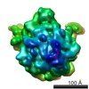
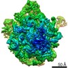
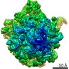
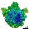
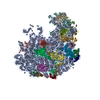
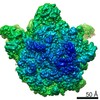
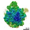
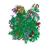
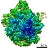
| タイトル | 50S ribosomal subunit assembly intermediate state 5 リボソーム リボソーム | |||||||||
 マップデータ マップデータ | ||||||||||
 試料 試料 |
| |||||||||
 キーワード キーワード | assembly precursor 48S 50S biogenesis in vitro reconstitution / RIBOSOME (リボソーム) | |||||||||
| 機能・相同性 |  機能・相同性情報transcriptional attenuation / endoribonuclease inhibitor activity / RNA-binding transcription regulator activity / positive regulation of ribosome biogenesis / negative regulation of cytoplasmic translation / DnaA-L2 complex / translation repressor activity / negative regulation of DNA-templated DNA replication initiation / ribosome assembly / mRNA regulatory element binding translation repressor activity ...transcriptional attenuation / endoribonuclease inhibitor activity / RNA-binding transcription regulator activity / positive regulation of ribosome biogenesis / negative regulation of cytoplasmic translation / DnaA-L2 complex / translation repressor activity / negative regulation of DNA-templated DNA replication initiation / ribosome assembly / mRNA regulatory element binding translation repressor activity / assembly of large subunit precursor of preribosome / response to reactive oxygen species / cytosolic ribosome assembly / regulation of cell growth / DNA-templated transcription termination / response to radiation / mRNA 5'-UTR binding / ribosomal large subunit assembly / large ribosomal subunit rRNA binding / ribosome binding / large ribosomal subunit / cytoplasmic translation / 5S rRNA binding / cytosolic large ribosomal subunit / transferase activity / tRNA binding / negative regulation of translation / rRNA binding / リボソーム / structural constituent of ribosome / 翻訳 (生物学) / response to antibiotic / mRNA binding / negative regulation of DNA-templated transcription / DNA binding / RNA binding / zinc ion binding / 細胞質基質 / 細胞質 機能・相同性情報transcriptional attenuation / endoribonuclease inhibitor activity / RNA-binding transcription regulator activity / positive regulation of ribosome biogenesis / negative regulation of cytoplasmic translation / DnaA-L2 complex / translation repressor activity / negative regulation of DNA-templated DNA replication initiation / ribosome assembly / mRNA regulatory element binding translation repressor activity ...transcriptional attenuation / endoribonuclease inhibitor activity / RNA-binding transcription regulator activity / positive regulation of ribosome biogenesis / negative regulation of cytoplasmic translation / DnaA-L2 complex / translation repressor activity / negative regulation of DNA-templated DNA replication initiation / ribosome assembly / mRNA regulatory element binding translation repressor activity / assembly of large subunit precursor of preribosome / response to reactive oxygen species / cytosolic ribosome assembly / regulation of cell growth / DNA-templated transcription termination / response to radiation / mRNA 5'-UTR binding / ribosomal large subunit assembly / large ribosomal subunit rRNA binding / ribosome binding / large ribosomal subunit / cytoplasmic translation / 5S rRNA binding / cytosolic large ribosomal subunit / transferase activity / tRNA binding / negative regulation of translation / rRNA binding / リボソーム / structural constituent of ribosome / 翻訳 (生物学) / response to antibiotic / mRNA binding / negative regulation of DNA-templated transcription / DNA binding / RNA binding / zinc ion binding / 細胞質基質 / 細胞質類似検索 - 分子機能 | |||||||||
| 生物種 |  Escherichia coli (大腸菌) / Escherichia coli K-12 (大腸菌) Escherichia coli (大腸菌) / Escherichia coli K-12 (大腸菌) | |||||||||
| 手法 | 単粒子再構成法 / クライオ電子顕微鏡法 / 解像度: 3.8 Å | |||||||||
 データ登録者 データ登録者 | Nikolay R / Hilal T | |||||||||
| 資金援助 |  ドイツ, 2件 ドイツ, 2件
| |||||||||
 引用 引用 | ジャーナル: Mol Cell / 年: 2018 タイトル: Structural Visualization of the Formation and Activation of the 50S Ribosomal Subunit during In Vitro Reconstitution. 著者: Rainer Nikolay / Tarek Hilal / Bo Qin / Thorsten Mielke / Jörg Bürger / Justus Loerke / Kathrin Textoris-Taube / Knud H Nierhaus / Christian M T Spahn / 要旨: The assembly of ribosomal subunits is an essential prerequisite for protein biosynthesis in all domains of life. Although biochemical and biophysical approaches have advanced our understanding of ...The assembly of ribosomal subunits is an essential prerequisite for protein biosynthesis in all domains of life. Although biochemical and biophysical approaches have advanced our understanding of ribosome assembly, our mechanistic comprehension of this process is still limited. Here, we perform an in vitro reconstitution of the Escherichia coli 50S ribosomal subunit. Late reconstitution products were subjected to high-resolution cryo-electron microscopy and multiparticle refinement analysis to reconstruct five distinct precursors of the 50S subunit with 4.3-3.8 Å resolution. These assembly intermediates define a progressive maturation pathway culminating in a late assembly particle, whose structure is more than 96% identical to a mature 50S subunit. Our structures monitor the formation and stabilization of structural elements in a nascent particle in unprecedented detail and identify the maturation of the rRNA-based peptidyl transferase center as the final critical step along the 50S assembly pathway. | |||||||||
| 履歴 |
|
- 構造の表示
構造の表示
| ムービー |
ムービービューア |
|---|---|
| 構造ビューア | EMマップ: SurfViewMolmilJmol/JSmol |
| 添付画像 |

- ダウンロードとリンク
-EMDBアーカイブ
| マップデータ | emd_4378.map.gz | 69.9 MB | EMDBマップデータ形式 | |
|---|---|---|---|---|
| ヘッダ (付随情報) | emd-4378-v30.xmlemd-4378.xml | 41.6 KB 41.6 KB | 表示 表示 | EMDBヘッダ |
| 画像 | 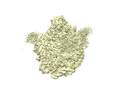 emd_4378.png emd_4378.png | 128.7 KB | ||
| Filedesc metadata | emd-4378.cif.gz | 9.6 KB | ||
| アーカイブディレクトリ |  http://ftp.pdbj.org/pub/emdb/structures/EMD-4378ftp://ftp.pdbj.org/pub/emdb/structures/EMD-4378 http://ftp.pdbj.org/pub/emdb/structures/EMD-4378ftp://ftp.pdbj.org/pub/emdb/structures/EMD-4378 | HTTPS FTP |
-関連構造データ
| 関連構造データ | 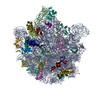 6gbzMC 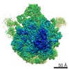 4379C 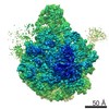 4380C 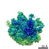 4381C 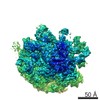 4382C 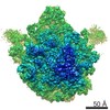 4383C 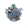 6gc0C 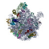 6gc4C 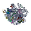 6gc6C  6gc7C 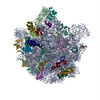 6gc8C M: このマップから作成された原子モデル C: 同じ文献を引用 ( |
|---|---|
| 類似構造データ |
-リンク
| EMDBのページ | EMDB (EBI/PDBe) / EMDataResource |
|---|---|
| 「今月の分子」の関連する項目 |


-マップ
| ファイル | ダウンロード / ファイル: emd_4378.map.gz / 形式: CCP4 / 大きさ: 75.1 MB / タイプ: IMAGE STORED AS FLOATING POINT NUMBER (4 BYTES) | ||||||||||||||||||||||||||||||||||||||||||||||||||||||||||||
|---|---|---|---|---|---|---|---|---|---|---|---|---|---|---|---|---|---|---|---|---|---|---|---|---|---|---|---|---|---|---|---|---|---|---|---|---|---|---|---|---|---|---|---|---|---|---|---|---|---|---|---|---|---|---|---|---|---|---|---|---|---|
| ボクセルのサイズ | X=Y=Z: 1.24 Å | ||||||||||||||||||||||||||||||||||||||||||||||||||||||||||||
| 密度 |
| ||||||||||||||||||||||||||||||||||||||||||||||||||||||||||||
| 対称性 | 空間群: 1 | ||||||||||||||||||||||||||||||||||||||||||||||||||||||||||||
| 詳細 | EMDB XML:
CCP4マップ ヘッダ情報:
| ||||||||||||||||||||||||||||||||||||||||||||||||||||||||||||
-添付データ
- 試料の構成要素
+全体 : 50S ribosomal subunit assembly intermediate state 5
+超分子 #1: 50S ribosomal subunit assembly intermediate state 5
+分子 #1: 23S ribosomal RNA
+分子 #28: 5S ribosomal RNA
+分子 #2: 50S ribosomal protein L2
+分子 #3: 50S ribosomal protein L3
+分子 #4: 50S ribosomal protein L4
+分子 #5: 50S ribosomal protein L5
+分子 #6: 50S ribosomal protein L6
+分子 #7: 50S ribosomal protein L9
+分子 #8: 50S ribosomal protein L13
+分子 #9: 50S ribosomal protein L14
+分子 #10: 50S ribosomal protein L15
+分子 #11: 50S ribosomal protein L17
+分子 #12: 50S ribosomal protein L18
+分子 #13: 50S ribosomal protein L19
+分子 #14: 50S ribosomal protein L20
+分子 #15: 50S ribosomal protein L21
+分子 #16: 50S ribosomal protein L22
+分子 #17: 50S ribosomal protein L23
+分子 #18: 50S ribosomal protein L24
+分子 #19: 50S ribosomal protein L25
+分子 #20: 50S ribosomal protein L27
+分子 #21: 50S ribosomal protein L28
+分子 #22: 50S ribosomal protein L29
+分子 #23: 50S ribosomal protein L30
+分子 #24: 50S ribosomal protein L32
+分子 #25: 50S ribosomal protein L33
+分子 #26: 50S ribosomal protein L34
+分子 #27: 50S ribosomal protein L35
+分子 #29: 50S ribosomal protein L16
-実験情報
-構造解析
| 手法 | クライオ電子顕微鏡法 |
|---|---|
 解析 解析 | 単粒子再構成法 |
| 試料の集合状態 | particle |
-試料調製
| 緩衝液 | pH: 7.6 |
|---|---|
| グリッド | モデル: Quantifoil R3/3 / 材質: COPPER / メッシュ: 300 / 前処理 - タイプ: GLOW DISCHARGE / 前処理 - 時間: 30 sec. |
| 凍結 | 凍結剤: ETHANE / 装置: FEI VITROBOT MARK I |
- 電子顕微鏡法
| 顕微鏡 | FEI POLARA 300 |
|---|---|
| 電子線 | 加速電圧: 300 kV / 電子線源: FIELD EMISSION GUN |
| 電子光学系 | 照射モード: FLOOD BEAM / 撮影モード: BRIGHT FIELDBright-field microscopy / Cs: 2.0 mm / 最大 デフォーカス(公称値): 5.0 µm / 最小 デフォーカス(公称値): 0.5 µm / 倍率(公称値): 31000 |
| 試料ステージ | ホルダー冷却材: NITROGEN |
| 撮影 | フィルム・検出器のモデル: GATAN K2 SUMMIT (4k x 4k) 検出モード: SUPER-RESOLUTION / デジタル化 - 画像ごとのフレーム数: 1-25 / 実像数: 1684 / 平均露光時間: 5.0 sec. / 平均電子線量: 25.0 e/Å2 |
| 実験機器 |  モデル: Tecnai Polara / 画像提供: FEI Company |
-画像解析
| 粒子像選択 | 選択した数: 154462 |
|---|---|
| 初期モデル | モデルのタイプ: OTHER 詳細: low-pass filtered reconstruction of bacterial 50S ribosomal subunit |
| 初期 角度割当 | タイプ: PROJECTION MATCHING / ソフトウェア - 名称: SPIDER (ver. 14) |
| 最終 3次元分類 | ソフトウェア - 名称: SPIDER (ver. 14) |
| 最終 角度割当 | タイプ: PROJECTION MATCHING / ソフトウェア - 名称: SPIDER (ver. 14) |
| 最終 再構成 | 想定した対称性 - 点群: C1 (非対称) / アルゴリズム: BACK PROJECTION / 解像度のタイプ: BY AUTHOR / 解像度: 3.8 Å / 解像度の算出法: FSC 0.143 CUT-OFF / ソフトウェア - 名称: SPIDER (ver. 14) / 使用した粒子像数: 51173 |
-原子モデル構築 1
| 精密化 | 空間: REAL / プロトコル: RIGID BODY FIT |
|---|---|
| 得られたモデル | PDB-6gbz: |
