National Institutes of Health/National Heart, Lung, and Blood Institute (NIH/NHLBI)
DP5-OD019800
米国
引用
ジャーナル: Nat Struct Mol Biol / 年: 2024 タイトル: Mechanism of autocatalytic activation during proteasome assembly. 著者: Benjamin Velez / Richard M Walsh / Shaun Rawson / Aida Razi / Lea Adams / Erignacio Fermin Perez / Fenglong Jiao / Marie Blickling / Tamayanthi Rajakumar / Darlene Fung / Lan Huang / John Hanna / 要旨: Many large molecular machines are too elaborate to assemble spontaneously and are built through ordered pathways orchestrated by dedicated chaperones. During assembly of the core particle (CP) of the ...Many large molecular machines are too elaborate to assemble spontaneously and are built through ordered pathways orchestrated by dedicated chaperones. During assembly of the core particle (CP) of the proteasome, where protein degradation occurs, its six active sites are simultaneously activated via cleavage of N-terminal propeptides. Such activation is autocatalytic and coupled to fusion of two half-CP intermediates, which protects cells by preventing activation until enclosure of the active sites within the CP interior. Here we uncover key mechanistic aspects of autocatalytic activation, which proceeds through alignment of the β5 and β2 catalytic triad residues, respectively, with these triads being misaligned before fusion. This mechanism contrasts with most other zymogens, in which catalytic centers are preformed. Our data also clarify the mechanism by which individual subunits can be added in a precise, temporally ordered manner. This work informs two decades-old mysteries in the proteasome field, with broader implications for protease biology and multisubunit complex assembly.
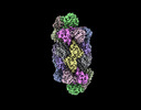
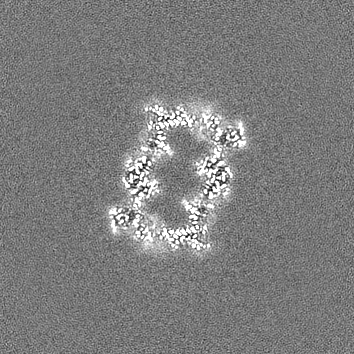
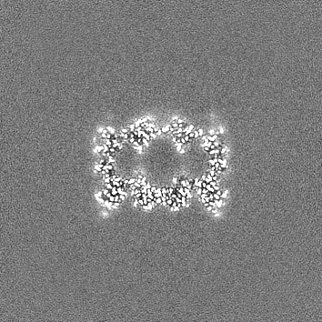
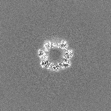
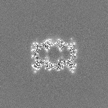
名称: Proteasome 20S Core Particle from Beta 3 D205 deletion mutant タイプ: complex / ID: 1 / 親要素: 0 / 含まれる分子: all 詳細: D205 is terminal most residue in the Beta 3 subunit. The deletion of this residue causes an accumulation of assembly intermediates including the Preholo-Proteasome. Mature 20S particle still forms.
 ムービー
ムービー コントローラー


 データを開く
 基本情報
基本情報
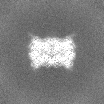
 マップデータ
 試料
試料 キーワード
キーワード Proteasome (プロテアソーム) /
  機能・相同性情報
 データ登録者
 米国, 1件
  引用
引用 構造の表示
構造の表示
 ダウンロードとリンク
 emd_41993.png
 http://ftp.pdbj.org/pub/emdb/structures/EMD-41993
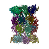


 Z
Z Y
Y X
X


 試料の構成要素
 解析
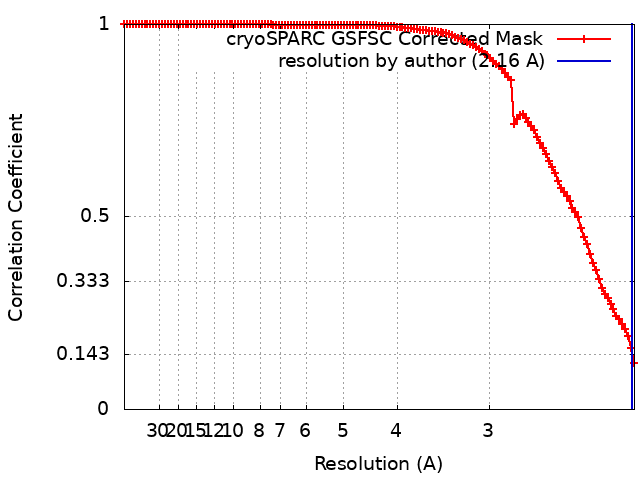
解析 電子顕微鏡法