 ムービー
ムービー コントローラー


+ データを開く

- 基本情報
基本情報
| 登録情報 | データベース: EMDB / ID: EMD-3418 | |||||||||
|---|---|---|---|---|---|---|---|---|---|---|
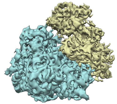
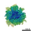
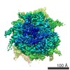
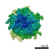
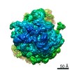
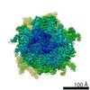
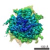
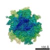
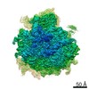
| タイトル | Subtomogram average of 80S ribosomes obtained using the VPP | |||||||||
 マップデータ マップデータ | Subtomogram average of the 80S ribosome obtained with the VPP | |||||||||
 試料 試料 |
| |||||||||
 キーワード キーワード |  Ribosome (リボソーム) / cryo-electron tomography / subtomogram averaging / phase plate Ribosome (リボソーム) / cryo-electron tomography / subtomogram averaging / phase plate | |||||||||
| 生物種 |  Oryctolagus cuniculus (ウサギ) Oryctolagus cuniculus (ウサギ) | |||||||||
| 手法 | サブトモグラム平均法 / クライオ電子顕微鏡法 / 解像度: 9.6 Å | |||||||||
 データ登録者 データ登録者 | Khoshouei M / Pfeffer S / Baumeister W / Foerster F / Danev R | |||||||||
 引用 引用 | ジャーナル: J Struct Biol / 年: 2017 タイトル: Subtomogram analysis using the Volta phase plate. 著者: Maryam Khoshouei / Stefan Pfeffer / Wolfgang Baumeister / Friedrich Förster / Radostin Danev /   要旨: Cryo-electron tomography (CET) and subtomogram analysis allow studying the structures of macromolecular complexes in their natural context. The radiation sensitivity of vitrified biological specimens ...Cryo-electron tomography (CET) and subtomogram analysis allow studying the structures of macromolecular complexes in their natural context. The radiation sensitivity of vitrified biological specimens and the resulting low signal-to-noise ratio (SNR) in CET limit the amount of structural information that can be mined from tomographic data. The Volta phase plate (VPP) has emerged as an effective means to increase the SNR and hence contrast compared to 'conventional' defocus-based phase contrast transmission electron microscopy (CTEM). Here, we assess the performance of the VPP compared to CTEM in subtomogram analysis, using the mammalian 80S ribosome as a test case. Accurate focusing is the major factor for achieving high resolution with the VPP, as highlighted by a comparison of slightly different focusing strategies. From only 1400 subtomograms, the VPP yields a subtomogram average of the mammalian 80S ribosome at 9.6Å resolution without laborious contrast transfer function (CTF) correction. The subtomogram averages obtained using CTEM approaches are comparable, but suffer from lower signal transfer in certain frequency bands due to the oscillations of the CTF. Our study demonstrates that the VPP is a valuable tool for subtomogram analysis, because it enables improved performance and efficiency in terms of structure localization and number of subtomograms required for a given resolution. | |||||||||
| 履歴 |
|
- 構造の表示
構造の表示
| ムービー |
 ムービービューア ムービービューア |
|---|---|
| 構造ビューア | EMマップ: SurfViewMolmilJmol/JSmol |
| 添付画像 |
- ダウンロードとリンク
-EMDBアーカイブ
| マップデータ | emd_3418.map.gz | 1.6 MB | EMDBマップデータ形式 | |
|---|---|---|---|---|
| ヘッダ (付随情報) | emd-3418-v30.xmlemd-3418.xml | 8.2 KB 8.2 KB | 表示 表示 | EMDBヘッダ |
| 画像 |  emd_3418.jpg emd_3418.jpg | 194.9 KB | ||
| アーカイブディレクトリ |  http://ftp.pdbj.org/pub/emdb/structures/EMD-3418ftp://ftp.pdbj.org/pub/emdb/structures/EMD-3418 http://ftp.pdbj.org/pub/emdb/structures/EMD-3418ftp://ftp.pdbj.org/pub/emdb/structures/EMD-3418 | HTTPS FTP |
-関連構造データ
| 関連構造データ |  3419C  3420C C: 同じ文献を引用 ( |
|---|---|
| 類似構造データ | |
| 電子顕微鏡画像生データ | EMPIAR-10064 (タイトル: VPP subtomogram averaging / Data size: 33.9 / Data #1: VPP_Ribosome, CTEM_Ribosome [class averages]) |
-リンク
| EMDBのページ | EMDB (EBI/PDBe) / EMDataResource |
|---|---|
| 「今月の分子」の関連する項目 |

-マップ
| ファイル | ダウンロード / ファイル: emd_3418.map.gz / 形式: CCP4 / 大きさ: 15.3 MB / タイプ: IMAGE STORED AS FLOATING POINT NUMBER (4 BYTES) | ||||||||||||||||||||||||||||||||||||||||||||||||||||||||||||
|---|---|---|---|---|---|---|---|---|---|---|---|---|---|---|---|---|---|---|---|---|---|---|---|---|---|---|---|---|---|---|---|---|---|---|---|---|---|---|---|---|---|---|---|---|---|---|---|---|---|---|---|---|---|---|---|---|---|---|---|---|---|
| 注釈 | Subtomogram average of the 80S ribosome obtained with the VPP | ||||||||||||||||||||||||||||||||||||||||||||||||||||||||||||
| ボクセルのサイズ | X=Y=Z: 2.62 Å | ||||||||||||||||||||||||||||||||||||||||||||||||||||||||||||
| 密度 |
| ||||||||||||||||||||||||||||||||||||||||||||||||||||||||||||
| 対称性 | 空間群: 1 | ||||||||||||||||||||||||||||||||||||||||||||||||||||||||||||
| 詳細 | EMDB XML:
CCP4マップ ヘッダ情報:
| ||||||||||||||||||||||||||||||||||||||||||||||||||||||||||||
-添付データ
- 試料の構成要素
-全体 : 80S ribosome
| 全体 | 名称: 80S ribosomeEukaryotic ribosome |
|---|---|
| 要素 |
|
-超分子 #1000: 80S ribosome
| 超分子 | 名称: 80S ribosome / タイプ: sample / ID: 1000 / Number unique components: 1 |
|---|
-超分子 #1: 80S ribosome
| 超分子 | 名称: 80S ribosome / タイプ: complex / ID: 1 / 組換発現: No / Ribosome-details: ribosome-eukaryote: ALL |
|---|---|
| 由来(天然) | 生物種: Oryctolagus cuniculus (ウサギ) / 別称: Rabbit / 細胞: Reticulocyte / 細胞中の位置: Cytoplasm |
-実験情報
-構造解析
| 手法 | クライオ電子顕微鏡法 |
|---|---|
 解析 解析 | サブトモグラム平均法 |
| 試料の集合状態 | particle |
-試料調製
| 緩衝液 | pH: 7.6 / 詳細: 20mM Hepes, 50mM KCl; 2mM MgCl2 |
|---|---|
| グリッド | 詳細: Quantifoil R 2/1 |
| 凍結 | 凍結剤: ETHANE-PROPANE MIXTURE / 装置: FEI VITROBOT MARK IV 手法: Blotting time of 3 seconds and a blot force of 0 before plunging |
- 電子顕微鏡法
| 顕微鏡 | FEI TITAN |
|---|---|
| 電子線 | 加速電圧: 300 kV / 電子線源: FIELD EMISSION GUN |
| 電子光学系 | 照射モード: FLOOD BEAM / 撮影モード: BRIGHT FIELDBright-field microscopy / 最大 デフォーカス(公称値): 0.0 µm / 最小 デフォーカス(公称値): 0.0 µm |
| 特殊光学系 | エネルギーフィルター - 名称: GIF Quantum エネルギーフィルター - エネルギー下限: 0.0 eV エネルギーフィルター - エネルギー上限: 20.0 eV |
| 試料ステージ | 試料ホルダーモデル: FEI TITAN KRIOS AUTOGRID HOLDER Tilt series - Axis1 - Min angle: -20 ° / Tilt series - Axis1 - Max angle: 20 ° |
| 日付 | 2015年5月6日 |
| 撮影 | カテゴリ: CCD フィルム・検出器のモデル: GATAN K2 SUMMIT (4k x 4k) 実像数: 80 / 平均電子線量: 30 e/Å2 |
-画像解析
| CTF補正 | 詳細: each tilt image |
|---|---|
| 最終 再構成 | 想定した対称性 - 点群: C1 (非対称) / 解像度のタイプ: BY AUTHOR / 解像度: 9.6 Å / 解像度の算出法: OTHER / ソフトウェア - 名称: TOM, AV3, PyTom / 使用したサブトモグラム数: 1400 |
