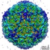
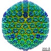
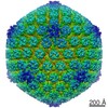
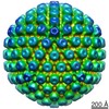
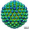
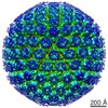
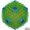
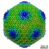
ジャーナル: J Virol / 年: 2016 タイトル: Internal Proteins of the Procapsid and Mature Capsids of Herpes Simplex Virus 1 Mapped by Bubblegram Imaging. 著者: Weimin Wu / William W Newcomb / Naiqian Cheng / Anastasia Aksyuk / Dennis C Winkler / Alasdair C Steven / 要旨: The herpes simplex virus 1 (HSV-1) capsid is a huge assembly, ∼1,250 Å in diameter, and is composed of thousands of protein subunits with a combined mass of ∼200 MDa, housing a 100-MDa genome. ...The herpes simplex virus 1 (HSV-1) capsid is a huge assembly, ∼1,250 Å in diameter, and is composed of thousands of protein subunits with a combined mass of ∼200 MDa, housing a 100-MDa genome. First, a procapsid is formed through coassembly of the surface shell with an inner scaffolding shell; then the procapsid matures via a major structural transformation, triggered by limited proteolysis of the scaffolding proteins. Three mature capsids are found in the nuclei of infected cells. A capsids are empty, B capsids retain a shrunken scaffolding shell, and C capsids-which develop into infectious virions-are filled with DNA and ostensibly have expelled the scaffolding shell. The possible presence of other internal proteins in C capsids has been moot as, in cryo-electron microscopy (cryo-EM), they would be camouflaged by the surrounding DNA. We have used bubblegram imaging to map internal proteins in all four capsids, aided by the discovery that the scaffolding protein is exceptionally prone to radiation-induced bubbling. We confirmed that this protein forms thick-walled inner shells in the procapsid and the B capsid. C capsids generate two classes of bubbles: one occupies positions beneath the vertices of the icosahedral surface shell, and the other is distributed throughout its interior. A likely candidate is the viral protease. A subpopulation of C capsids bubbles particularly profusely and may represent particles in which expulsion of scaffold and DNA packaging are incomplete. Based on the procapsid structure, we propose that the axial channels of hexameric capsomers afford the pathway via which the scaffolding protein is expelled. IMPORTANCE: In addition to DNA, capsids of tailed bacteriophages and their distant relatives, herpesviruses, contain internal proteins. These proteins are often essential for infectivity but are ...IMPORTANCE: In addition to DNA, capsids of tailed bacteriophages and their distant relatives, herpesviruses, contain internal proteins. These proteins are often essential for infectivity but are difficult to locate within the virion. A novel adaptation of cryo-EM based on detecting gas bubbles generated by radiation damage was used to localize internal proteins of HSV-1, yielding insights into how capsid maturation is regulated. The scaffolding protein, which forms inner shells in the procapsid and B capsid, is exceptionally bubbling-prone. In the mature DNA-filled C capsid, a previously undetected protein was found to underlie the icosahedral vertices: this is tentatively assigned as a storage form of the viral protease. We also observed a capsid species that appears to contain substantial amounts of scaffolding protein as well as DNA, suggesting that DNA packaging and expulsion of the scaffolding protein are coupled processes.
カテゴリ: FILM / フィルム・検出器のモデル: KODAK SO-163 FILM デジタル化 - スキャナー: NIKON SUPER COOLSCAN 9000 デジタル化 - サンプリング間隔: 6.35 µm / 実像数: 18 / 平均電子線量: 105 e/Å2
-
画像解析
CTF補正
詳細: average of particles
最終 再構成
想定した対称性 - 点群: I (正20面体型対称) / アルゴリズム: OTHER / 解像度のタイプ: BY AUTHOR / 解像度: 25.0 Å / 解像度の算出法: OTHER / ソフトウェア - 名称: EMAN / 詳細: We are not aiming at resolution. / 使用した粒子像数: 235
 ムービー
ムービー コントローラー


 データを開く
 基本情報
基本情報 マップデータ
 試料
試料 キーワード
キーワード Herpes Simplex Virus (単純ヘルペスウイルス) /

 データ登録者
 引用
引用
 構造の表示
構造の表示 ムービービューア
 ダウンロードとリンク
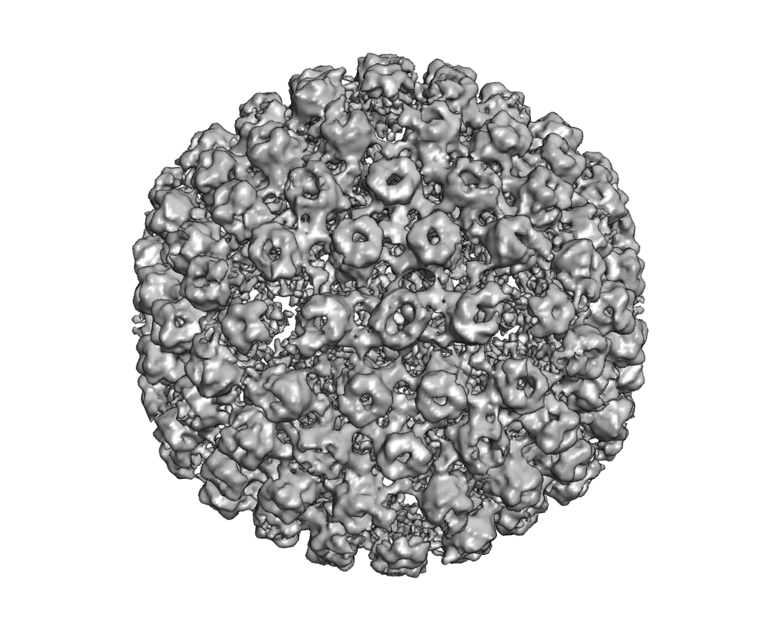 EMD-3358.png
 http://ftp.pdbj.org/pub/emdb/structures/EMD-3358
 試料の構成要素


 解析
解析 電子顕微鏡法