 ムービー
ムービー コントローラー


+ データを開く

- 基本情報
基本情報
| 登録情報 | データベース: EMDB / ID: EMD-3215 | |||||||||
|---|---|---|---|---|---|---|---|---|---|---|
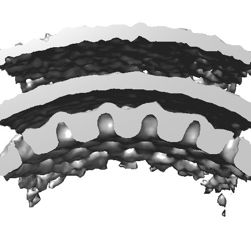
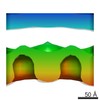
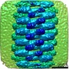
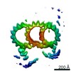
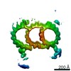
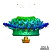
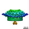
| タイトル | Sub-tomogram averaging of electron cryo-microscopic data taken from focused-ion beam milled lamellae of nuclei of Pseudorabies virus (PrV) nuclear egress complex-expressing cells | |||||||||
 マップデータ マップデータ | Sub-tomogram average of nuclear egress complex from vesicles observed inside nuclei of cells co-expressing PrV UL31 and UL34 by focus-ion beam milling and electron cryo-tomography. | |||||||||
 試料 試料 |
| |||||||||
 キーワード キーワード |  alphaherpesvirinae (アルファヘルペスウイルス亜科) / herpesvirus simplex / HSV-1 (単純ヘルペスウイルス) / pseudorabies virus (オーエスキー病) / PrV / nuclear egress complex / nuclear envelope (核膜) / nucleoplasmic reticulum / inner nuclear membrane (核膜) / UL31 / UL34 / vesicle transport (小胞) / nucleo-cytoplasmic transport / nanovesicles / cryoEM (低温電子顕微鏡法) / cryoET / electron cryo-microscopy (低温電子顕微鏡法) / electron cryo-tomography / cryoFIB / focused ion beam milling / FIB-SEM alphaherpesvirinae (アルファヘルペスウイルス亜科) / herpesvirus simplex / HSV-1 (単純ヘルペスウイルス) / pseudorabies virus (オーエスキー病) / PrV / nuclear egress complex / nuclear envelope (核膜) / nucleoplasmic reticulum / inner nuclear membrane (核膜) / UL31 / UL34 / vesicle transport (小胞) / nucleo-cytoplasmic transport / nanovesicles / cryoEM (低温電子顕微鏡法) / cryoET / electron cryo-microscopy (低温電子顕微鏡法) / electron cryo-tomography / cryoFIB / focused ion beam milling / FIB-SEM | |||||||||
| 生物種 |  Sus scrofa (ブタ) Sus scrofa (ブタ) | |||||||||
| 手法 | サブトモグラム平均法 / クライオ電子顕微鏡法 / ネガティブ染色法 / 解像度: 35.0 Å | |||||||||
 データ登録者 データ登録者 | Hagen C / Siebert CA / Dent KC / Vasishtan D / Zeev Ben Mordehai T / Grange M / Klupp BG / Mettenleiter T / Gruenewald K | |||||||||
 引用 引用 | ジャーナル: Cell / 年: 2015 タイトル: Structural Basis of Vesicle Formation at the Inner Nuclear Membrane. 著者: Christoph Hagen / Kyle C Dent / Tzviya Zeev-Ben-Mordehai / Michael Grange / Jens B Bosse / Cathy Whittle / Barbara G Klupp / C Alistair Siebert / Daven Vasishtan / Felix J B Bäuerlein / ...著者: Christoph Hagen / Kyle C Dent / Tzviya Zeev-Ben-Mordehai / Michael Grange / Jens B Bosse / Cathy Whittle / Barbara G Klupp / C Alistair Siebert / Daven Vasishtan / Felix J B Bäuerlein / Juliana Cheleski / Stephan Werner / Peter Guttmann / Stefan Rehbein / Katja Henzler / Justin Demmerle / Barbara Adler / Ulrich Koszinowski / Lothar Schermelleh / Gerd Schneider / Lynn W Enquist / Jürgen M Plitzko / Thomas C Mettenleiter / Kay Grünewald /    要旨: Vesicular nucleo-cytoplasmic transport is becoming recognized as a general cellular mechanism for translocation of large cargoes across the nuclear envelope. Cargo is recruited, enveloped at the ...Vesicular nucleo-cytoplasmic transport is becoming recognized as a general cellular mechanism for translocation of large cargoes across the nuclear envelope. Cargo is recruited, enveloped at the inner nuclear membrane (INM), and delivered by membrane fusion at the outer nuclear membrane. To understand the structural underpinning for this trafficking, we investigated nuclear egress of progeny herpesvirus capsids where capsid envelopment is mediated by two viral proteins, forming the nuclear egress complex (NEC). Using a multi-modal imaging approach, we visualized the NEC in situ forming coated vesicles of defined size. Cellular electron cryo-tomography revealed a protein layer showing two distinct hexagonal lattices at its membrane-proximal and membrane-distant faces, respectively. NEC coat architecture was determined by combining this information with integrative modeling using small-angle X-ray scattering data. The molecular arrangement of the NEC establishes the basic mechanism for budding and scission of tailored vesicles at the INM. #1: ジャーナル: Cell Reports / 年: 2015タイトル: Crystal structure of the herpesvirus nuclear egress complex provides insights into inner nuclear membrane remodelling 著者: Zeev-Ben-Mordehai T / Weberruss M / Lorenz M / Cheleski J / Hellberg T / Whittle C / El Omari K / Vasishtan D / Dent KC Harlos K / Franzke K / Hagen C / Klupp B / Antonin W / Mettenleiter TC / Gruenewald K | |||||||||
| 履歴 |
|
- 構造の表示
構造の表示
| ムービー |
 ムービービューア ムービービューア |
|---|---|
| 構造ビューア | EMマップ: SurfViewMolmilJmol/JSmol |
| 添付画像 |
- ダウンロードとリンク
-EMDBアーカイブ
| マップデータ | emd_3215.map.gz | 948.9 KB | EMDBマップデータ形式 | |
|---|---|---|---|---|
| ヘッダ (付随情報) | emd-3215-v30.xmlemd-3215.xml | 13.7 KB 13.7 KB | 表示 表示 | EMDBヘッダ |
| 画像 |  emd_3215.png emd_3215.png | 67.8 KB | ||
| アーカイブディレクトリ |  http://ftp.pdbj.org/pub/emdb/structures/EMD-3215ftp://ftp.pdbj.org/pub/emdb/structures/EMD-3215 http://ftp.pdbj.org/pub/emdb/structures/EMD-3215ftp://ftp.pdbj.org/pub/emdb/structures/EMD-3215 | HTTPS FTP |
-関連構造データ
-リンク
| EMDBのページ | EMDB (EBI/PDBe) / EMDataResource |
|---|
-マップ
| ファイル | ダウンロード / ファイル: emd_3215.map.gz / 形式: CCP4 / 大きさ: 1001 KB / タイプ: IMAGE STORED AS FLOATING POINT NUMBER (4 BYTES) | ||||||||||||||||||||||||||||||||||||||||||||||||||||||||||||
|---|---|---|---|---|---|---|---|---|---|---|---|---|---|---|---|---|---|---|---|---|---|---|---|---|---|---|---|---|---|---|---|---|---|---|---|---|---|---|---|---|---|---|---|---|---|---|---|---|---|---|---|---|---|---|---|---|---|---|---|---|---|
| 注釈 | Sub-tomogram average of nuclear egress complex from vesicles observed inside nuclei of cells co-expressing PrV UL31 and UL34 by focus-ion beam milling and electron cryo-tomography. | ||||||||||||||||||||||||||||||||||||||||||||||||||||||||||||
| ボクセルのサイズ | X=Y=Z: 11.4 Å | ||||||||||||||||||||||||||||||||||||||||||||||||||||||||||||
| 密度 |
| ||||||||||||||||||||||||||||||||||||||||||||||||||||||||||||
| 対称性 | 空間群: 1 | ||||||||||||||||||||||||||||||||||||||||||||||||||||||||||||
| 詳細 | EMDB XML:
CCP4マップ ヘッダ情報:
| ||||||||||||||||||||||||||||||||||||||||||||||||||||||||||||
-添付データ
- 試料の構成要素
-全体 : porcine epithelial-like embryonic EFN-R kidney cells stably co-ex...
| 全体 | 名称: porcine epithelial-like embryonic EFN-R kidney cells stably co-expressing PrV UL31 and UL34, the latter fused with GFP (cell line designated as BK/EF/UL31/34gfp catalogue No. RIE 1083 of the ...名称: porcine epithelial-like embryonic EFN-R kidney cells stably co-expressing PrV UL31 and UL34, the latter fused with GFP (cell line designated as BK/EF/UL31/34gfp catalogue No. RIE 1083 of the Collection of Cell Lines in Veterinary Medicine at the FLI, Greifswald-Insel Riems, Germany) |
|---|---|
| 要素 |
|
-超分子 #1000: porcine epithelial-like embryonic EFN-R kidney cells stably co-ex...
| 超分子 | 名称: porcine epithelial-like embryonic EFN-R kidney cells stably co-expressing PrV UL31 and UL34, the latter fused with GFP (cell line designated as BK/EF/UL31/34gfp catalogue No. RIE 1083 of the ...名称: porcine epithelial-like embryonic EFN-R kidney cells stably co-expressing PrV UL31 and UL34, the latter fused with GFP (cell line designated as BK/EF/UL31/34gfp catalogue No. RIE 1083 of the Collection of Cell Lines in Veterinary Medicine at the FLI, Greifswald-Insel Riems, Germany) タイプ: sample / ID: 1000 / 詳細: cellular 集合状態: coat of ~500 hexamers of pUL31/pUL34 heterodimers per vesicle Number unique components: 2 |
|---|
-超分子 #1: Nuclear envelope
| 超分子 | 名称: Nuclear envelope / タイプ: organelle_or_cellular_component / ID: 1 / Name.synonym: Inner and outer nuclear membranes 詳細: two proteins (pUL31 and pUL34) of pseudorabies virus (PrV) are co-expressed in the cells forming there the herpesviral nuclear egress complex lining as a coat perinuclear vesicles 集合状態: heterodimer / 組換発現: No |
|---|---|
| Ref INTERPRO | divclassse qspanoncli ckpopupspa nclassgree n(this)spandata popltspanc lassquotlo adingbarqu otgtltimgs rcquotimgl oadinggifq uotdecodin gquotasync quotgtltsp angtdataur lajaxphp?m odetaxoamp ... divclassse qspanoncli ckpopupspa nclassgree n(this)spandata popltspanc lassquotlo adingbarqu otgtltimgs rcquotimgl oadinggifq uotdecodin gquotasync quotgtltsp angtdataur lajaxphp?m odetaxoamp kIPR021152 ampajax1cl asspoptrgi IPR021152i spandiv |
| Ref INTERPRO | divclassse qspanoncli ckpopupspa nclassgree n(this)spandata popltspanc lassquotlo adingbarqu otgtltimgs rcquotimgl oadinggifq uotdecodin gquotasync quotgtltsp angtdataur lajaxphp?m odetaxoamp ... divclassse qspanoncli ckpopupspa nclassgree n(this)spandata popltspanc lassquotlo adingbarqu otgtltimgs rcquotimgl oadinggifq uotdecodin gquotasync quotgtltsp angtdataur lajaxphp?m odetaxoamp kIPR007626 ampajax1cl asspoptrgi IPR007626i spandiv |
| 由来(天然) | 生物種: Sus scrofa (ブタ) / 別称: Pig / 組織: Kidney / 細胞: Epithelial-like embryonic / Organelle: Nucleus / 細胞中の位置: Nuclear mebranes |
| 分子量 | 実験値: 60 KDa / 理論値: 60 KDa |
-実験情報
-構造解析
| 手法 | ネガティブ染色法, クライオ電子顕微鏡法 |
|---|---|
 解析 解析 | サブトモグラム平均法 |
| 試料の集合状態 | cell |
-試料調製
| 染色 | タイプ: NEGATIVE / 詳細: no staining |
|---|---|
| グリッド | 詳細: glow discharged standard 3.05 mm electron microscopy 200 mesh gold grids covered with a perforated carbon foil (R2/1; Quantifoil Micro Tools GmbH, Jena, Germany); focused-ion beam milled lamella |
| 凍結 | 凍結剤: ETHANE-PROPANE MIXTURE / 装置: HOMEMADE PLUNGER Timed resolved state: two days of incubation (37 degree C, 5 % CO2) in plastic microscope slide growth chambers (mue-slide 2x9 well; Ibidi GmbH) before cryo-immobilization 手法: blotted manually with a bent strip of Whatman No. 1 filter paper from the non-coated grid side for 2 to 3 s immediately before vitrification by the gravity-driven plunging apparatus in a ...手法: blotted manually with a bent strip of Whatman No. 1 filter paper from the non-coated grid side for 2 to 3 s immediately before vitrification by the gravity-driven plunging apparatus in a ethane/propane mixture cooled by liquid nitrogen |
- 電子顕微鏡法
| 顕微鏡 | FEI POLARA 300 |
|---|---|
| 電子線 | 加速電圧: 300 kV / 電子線源: FIELD EMISSION GUN |
| 電子光学系 | 倍率(補正後): 52650 / 照射モード: FLOOD BEAM / 撮影モード: BRIGHT FIELDBright-field microscopy / Cs: 2 mm / 最大 デフォーカス(公称値): -6.0 µm / 最小 デフォーカス(公称値): -6.0 µm / 倍率(公称値): 22500 |
| 特殊光学系 | エネルギーフィルター - 名称: Gatan GIF 2 エネルギーフィルター - エネルギー下限: 0.0 eV エネルギーフィルター - エネルギー上限: 20.0 eV |
| 試料ステージ | 試料ホルダーモデル: OTHER / Tilt series - Axis1 - Min angle: -50 ° / Tilt series - Axis1 - Max angle: 52 ° |
| 詳細 | 2048 x 2048 |
| 日付 | 2013年4月26日 |
| 撮影 | カテゴリ: CCD / フィルム・検出器のモデル: GATAN MULTISCAN / 実像数: 35 / 平均電子線量: 114 e/Å2 詳細: electron cryo-tomographic tilt series with 3 degree spacing ビット/ピクセル: 32 |
| 実験機器 |  モデル: Tecnai Polara / 画像提供: FEI Company |
-画像解析
| 最終 再構成 | 想定した対称性 - 点群: C1 (非対称) / アルゴリズム: OTHER / 解像度のタイプ: BY AUTHOR / 解像度: 35.0 Å / 解像度の算出法: OTHER / ソフトウェア - 名称: IMOD / 使用したサブトモグラム数: 300 |
|---|---|
| 詳細 | 'Particle' positions and orientations were initially approximated by modelling intraluminal vesicles as a set of points distributed over a sphere. After orienting particles to align their primary axes (defined as normal to the vesicle membrane) to the Y-axis, iterative 3D orientation search and translational alignment was carried out using PEET according to standard methods. Particles from each vesicle were aligned and average independently. For further information please refer to the primary citation. |
