Ministry of Education, Culture, Sports, Science and Technology (Japan)
JP17H05825
日本
Ministry of Education, Culture, Sports, Science and Technology (Japan)
JP19H04845
日本
Japan Society for the Promotion of Science (JSPS)
20H03078
日本
Japan Agency for Medical Research and Development (AMED)
JP18am0101072
日本
引用
ジャーナル: J Virol / 年: 2022 タイトル: Particle Morphology of Medusavirus Inside and Outside the Cells Reveals a New Maturation Process of Giant Viruses. 著者: Ryoto Watanabe / Chihong Song / Yoko Kayama / Masaharu Takemura / Kazuyoshi Murata / 要旨: Medusavirus, a giant virus, is phylogenetically closer to eukaryotes than the other giant viruses and has been recently classified as an independent species. However, details of its morphology and ...Medusavirus, a giant virus, is phylogenetically closer to eukaryotes than the other giant viruses and has been recently classified as an independent species. However, details of its morphology and maturation process in host cells remain unclear. Here, we investigated the particle morphology of medusavirus inside and outside infected cells using conventional transmission electron microscopy (C-TEM) and cryo-electron microscopy (cryo-EM). The C-TEM of amoebae infected with the medusavirus showed four types of particles, i.e., pseudo-DNA-empty (p-Empty), DNA-empty (Empty), semi-DNA-full (s-Full), and DNA-full (Full). Time-dependent changes in the four types of particles and their intracellular localization suggested a new maturation process for the medusavirus. Viral capsids and viral DNAs are produced independently in the cytoplasm and nucleus, respectively, and only the empty particles located near the host nucleus can incorporate the viral DNA into the capsid. Therefore, all four types of particles were found outside the cells. The cryo-EM of these particles showed that the intact virus structure, covered with three different types of spikes, was preserved among all particle types, although with minor size-related differences. The internal membrane exhibited a structural array similar to that of the capsid, interacted closely with the capsid, and displayed open membrane structures in the Empty and p-Empty particles. The results suggest that these open structures in the internal membrane are used for an exchange of scaffold proteins and viral DNA during the maturation process. This new model of the maturation process of medusavirus provides insight into the structural and behavioral diversity of giant viruses. Giant viruses exhibit diverse morphologies and maturation processes. In this study, medusavirus showed four types of particle morphologies, both inside and outside the infected cells, when propagated in amoeba culture. Time-course analysis and intracellular localization of the medusavirus in the infected cells suggested a new maturation process via the four types of particles. Like the previously reported pandoravirus, the viral DNA of medusavirus is replicated in the host's nucleus. However, viral capsids are produced independently in the host cytoplasm, and only empty capsids near the nucleus can take up viral DNA. As a result, many immature particles were released from the host cell along with the mature particles. The capsid structure is well conserved among the four types of particles, except for the open membrane structures in the empty particles, suggesting that they are used to exchange scaffold proteins for viral DNAs. These findings indicate that medusavirus has a unique maturation process.
 ムービー
ムービー コントローラー


 データを開く
 基本情報
基本情報
 マップデータ
 試料
試料 キーワード
キーワード Giant virus / NCLDV /
  Acanthamoeba castellanii medusavirus (ウイルス)
 データ登録者
 日本, 4件
  引用
引用 構造の表示
構造の表示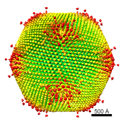
 ダウンロードとリンク
 EMDBマップデータ形式
 emd_32073.png
 http://ftp.pdbj.org/pub/emdb/structures/EMD-32073
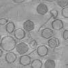
 試料の構成要素
 Acanthamoeba castellanii (カステラーニアメーバ)
 解析
解析 電子顕微鏡法

