primary metabolic process / large ribosomal subunit rRNA binding / large ribosomal subunit / regulation of translation / small ribosomal subunit / 5S rRNA binding / transferase activity / tRNA binding / rRNA binding / リボソーム ...primary metabolic process / large ribosomal subunit rRNA binding / large ribosomal subunit / regulation of translation / small ribosomal subunit / 5S rRNA binding / transferase activity / tRNA binding / rRNA binding / リボソーム / structural constituent of ribosome / 翻訳 (生物学) / ribonucleoprotein complex / mRNA binding / zinc ion binding / 細胞質基質 / 細胞質 類似検索 - 分子機能
Ribosomal protein L31 type B / Ribosome hibernation promoting factor/RaiA / Ribosome hibernation promotion factor-like / Sigma 54 modulation protein / S30EA ribosomal protein / Ribosomal protein L1, bacterial-type / Ribosomal protein L10, eubacterial, conserved site / Ribosomal protein L10 signature. / Ribosomal protein L25, long-form / Ribosomal protein L25, beta domain / Ribosomal protein L25, C-terminal ...Ribosomal protein L31 type B / Ribosome hibernation promoting factor/RaiA / Ribosome hibernation promotion factor-like / Sigma 54 modulation protein / S30EA ribosomal protein / Ribosomal protein L1, bacterial-type / Ribosomal protein L10, eubacterial, conserved site / Ribosomal protein L10 signature. / Ribosomal protein L25, long-form / Ribosomal protein L25, beta domain / Ribosomal protein L25, C-terminal / Ribosomal protein TL5, C-terminal domain / Ribosomal protein L10 / : / Ribosomal protein S14, type Z / Ribosomal protein L1, conserved site / Ribosomal protein L1 / Ribosomal protein L1 signature. / Ribosomal protein L1, 3-layer alpha/beta-sandwich / Ribosomal protein S21, conserved site / Ribosomal protein S21 signature. / Ribosomal protein L1-like / Ribosomal protein L1/ribosomal biogenesis protein / Ribosomal protein L1p/L10e family / Ribosomal protein L11, bacterial-type / Ribosomal protein S21 superfamily / Ribosomal protein S21 / Ribosomal protein S16, conserved site / Ribosomal protein S16 signature. / Ribosomal protein S21 / Ribosomal protein L31 signature. / Ribosomal protein L31 / Ribosomal protein L31 superfamily / Ribosomal protein L31 / Ribosomal protein L11, conserved site / Ribosomal protein L10-like domain superfamily / Ribosomal protein L21, conserved site / Ribosomal protein L21 signature. / Ribosomal protein L11 signature. / Ribosomal protein L10P / Ribosomal protein L10 / Ribosomal protein L16 signature 1. / : / Ribosomal protein L6, conserved site / Ribosomal protein L6 signature 1. / Ribosomal protein L16, conserved site / Ribosomal protein L16 signature 2. / Ribosomal protein L11, N-terminal / Ribosomal protein L9 signature. / Ribosomal protein L17 signature. / Ribosomal protein L9, bacteria/chloroplast / Ribosomal protein L9, C-terminal / Ribosomal protein L9, C-terminal domain / Ribosomal protein L9, C-terminal domain superfamily / Ribosomal protein L11/L12 / Ribosomal protein L11, C-terminal / Ribosomal protein L11, C-terminal domain superfamily / Ribosomal protein L11/L12, N-terminal domain superfamily / Ribosomal protein L11/L12 / Ribosomal protein L11, N-terminal domain / Ribosomal protein L11, RNA binding domain / Ribosomal L25p family / Ribosomal protein L25 / Ribosomal protein S14/S29 / Ribosomal protein L28/L24 superfamily / Ribosomal protein L36 signature. / Ribosomal protein L25/Gln-tRNA synthetase, N-terminal / Ribosomal protein L32p, bacterial type / Ribosomal protein L25/Gln-tRNA synthetase, anti-codon-binding domain superfamily / Ribosomal protein L9, N-terminal domain superfamily / Ribosomal protein L9 / Ribosomal protein L9, N-terminal / Ribosomal protein L9, N-terminal domain / Ribosomal protein L28 / Ribosomal protein L35, conserved site / Ribosomal protein L35 signature. / Ribosomal protein L33, conserved site / Ribosomal protein L33 signature. / Ribosomal protein L35, non-mitochondrial / Ribosomal protein L5, bacterial-type / Ribosomal protein L6, bacterial-type / Ribosomal protein L18, bacterial-type / Ribosomal protein L19, conserved site / Ribosomal protein L19 signature. / Ribosomal protein L36 / Ribosomal protein L36 superfamily / Ribosomal protein L36 / Ribosomal protein L9/RNase H1, N-terminal / Ribosomal protein L20 signature. / Ribosomal protein S3, bacterial-type / Ribosomal protein L27, conserved site / Ribosomal protein L27 signature. / Ribosomal protein S19, bacterial-type / Ribosomal protein S7, bacterial/organellar-type / Ribosomal protein S11, bacterial-type / Ribosomal protein S13, bacterial-type / Ribosomal protein S20 / Ribosomal protein S20 superfamily / Ribosomal protein S20 / Ribosomal protein S9, bacterial/plastid / Ribosomal protein L14P, bacterial-type 類似検索 - ドメイン・相同性
Large ribosomal subunit protein bL9 / Small ribosomal subunit protein bS18 / Small ribosomal subunit protein bS6 / Uncharacterized protein / Large ribosomal subunit protein bL20 / Large ribosomal subunit protein bL35 / Large ribosomal subunit protein bL31B / Small ribosomal subunit protein bS21 / Small ribosomal subunit protein uS9 / Large ribosomal subunit protein uL13 ...Large ribosomal subunit protein bL9 / Small ribosomal subunit protein bS18 / Small ribosomal subunit protein bS6 / Uncharacterized protein / Large ribosomal subunit protein bL20 / Large ribosomal subunit protein bL35 / Large ribosomal subunit protein bL31B / Small ribosomal subunit protein bS21 / Small ribosomal subunit protein uS9 / Large ribosomal subunit protein uL13 / Large ribosomal subunit protein bL28 / Small ribosomal subunit protein uS7 / Small ribosomal subunit protein uS12 / Large ribosomal subunit protein uL10 / Large ribosomal subunit protein uL1 / Large ribosomal subunit protein uL11 / Large ribosomal subunit protein bL33 / Ribosomal subunit interface protein / Large ribosomal subunit protein uL16 / Large ribosomal subunit protein uL29 / Small ribosomal subunit protein uS17 / Large ribosomal subunit protein uL14 / Large ribosomal subunit protein uL24 / Large ribosomal subunit protein uL5 / Small ribosomal subunit protein uS14 / Small ribosomal subunit protein uS8 / Large ribosomal subunit protein uL6 / Large ribosomal subunit protein uL18 / Small ribosomal subunit protein uS5 / Large ribosomal subunit protein uL15 / Large ribosomal subunit protein bL36 / Small ribosomal subunit protein uS13 / Small ribosomal subunit protein uS11 / Large ribosomal subunit protein bL17 / Small ribosomal subunit protein uS4 / Small ribosomal subunit protein bS16 / Large ribosomal subunit protein bL19 / Large ribosomal subunit protein bL32 / Large ribosomal subunit protein bL21 / Large ribosomal subunit protein bL27 / Large ribosomal subunit protein bL25 / Small ribosomal subunit protein uS15 / Large ribosomal subunit protein bL34 / Small ribosomal subunit protein bS20 / Small ribosomal subunit protein uS10 / Large ribosomal subunit protein uL3 / Large ribosomal subunit protein uL4 / Large ribosomal subunit protein uL23 / Large ribosomal subunit protein uL2 / Small ribosomal subunit protein uS19 / Large ribosomal subunit protein uL22 / Small ribosomal subunit protein uS3 類似検索 - 構成要素
National Institutes of Health/National Institute of General Medical Sciences (NIH/NIGMS)
R01 GM61576
米国
引用
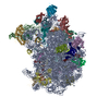
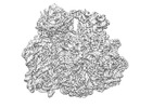
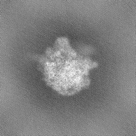
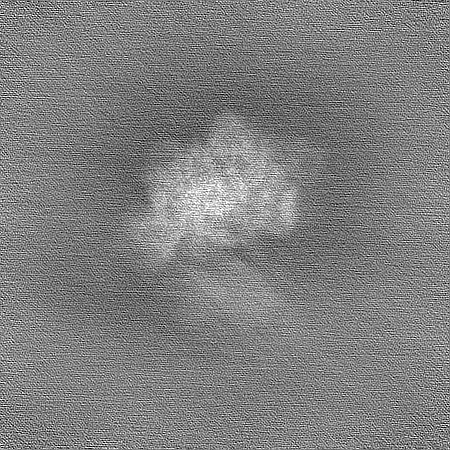
ジャーナル: Nat Commun / 年: 2023 タイトル: The structure of a hibernating ribosome in a Lyme disease pathogen. 著者: Manjuli R Sharma / Swati R Manjari / Ekansh K Agrawal / Pooja Keshavan / Ravi K Koripella / Soneya Majumdar / Ashley L Marcinkiewicz / Yi-Pin Lin / Rajendra K Agrawal / Nilesh K Banavali / 要旨: The spirochete bacterial pathogen Borrelia (Borreliella) burgdorferi (Bbu) affects more than 10% of the world population and causes Lyme disease in about half a million people in the US annually. ...The spirochete bacterial pathogen Borrelia (Borreliella) burgdorferi (Bbu) affects more than 10% of the world population and causes Lyme disease in about half a million people in the US annually. Therapy for Lyme disease includes antibiotics that target the Bbu ribosome. Here we present the structure of the Bbu 70S ribosome obtained by single particle cryo-electron microscopy at 2.9 Å resolution, revealing a bound hibernation promotion factor protein and two genetically non-annotated ribosomal proteins bS22 and bL38. The ribosomal protein uL30 in Bbu has an N-terminal α-helical extension, partly resembling the mycobacterial bL37 protein, suggesting evolution of bL37 and a shorter uL30 from a longer uL30 protein. Its analogy to proteins uL30m and mL63 in mammalian mitochondrial ribosomes also suggests a plausible evolutionary pathway for expansion of protein content in mammalian mitochondrial ribosomes. Computational binding free energy predictions for antibiotics reflect subtle distinctions in antibiotic-binding sites in the Bbu ribosome. Discovery of these features in the Bbu ribosome may enable better ribosome-targeted antibiotic design for Lyme disease treatment.
 ムービー
ムービー コントローラー


 データを開く
 基本情報
基本情報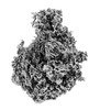
 マップデータ
 試料
試料 キーワード
キーワード bacterial (細菌) /
  機能・相同性情報
 データ登録者
 米国, 1件
  引用
引用 構造の表示
構造の表示
 ダウンロードとリンク
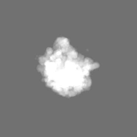
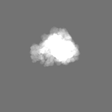
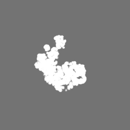
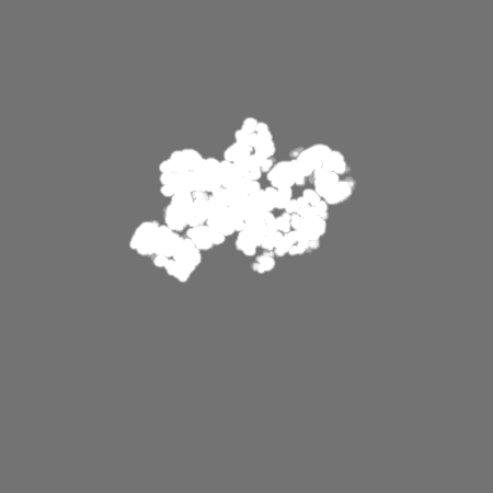
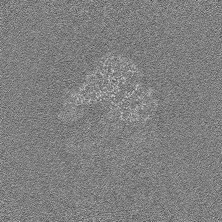
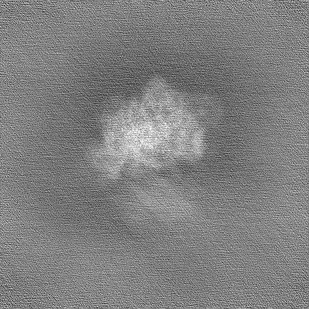
 emd_29304.png
 http://ftp.pdbj.org/pub/emdb/structures/EMD-29304
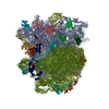


 Z
Z Y
Y X
X


 試料の構成要素
 解析
解析 電子顕微鏡法

