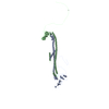
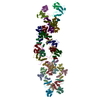
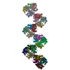
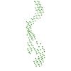
ジャーナル: J Mol Biol / 年: 2014 タイトル: Dynactin 3D structure: implications for assembly and dynein binding. 著者: Hiroshi Imai / Akihiro Narita / Yuichiro Maéda / Trina A Schroer / 要旨: The multisubunit protein complex, dynactin, is an essential component of the cytoplasmic dynein motor. High-resolution structural work on dynactin and the dynein/dynactin supercomplex has been ...The multisubunit protein complex, dynactin, is an essential component of the cytoplasmic dynein motor. High-resolution structural work on dynactin and the dynein/dynactin supercomplex has been limited to small subunits and recombinant fragments that do not report fully on either ≈1MDa assembly. In the present study, we used negative-stain electron microscopy and image analysis based on random conical tilt reconstruction to obtain a three-dimensional (3D) structure of native vertebrate dynactin. The 35-nm-long dynactin molecule has a V-shaped shoulder at one end and a flattened tip at the other end, both offset relative to the long axis of the actin-related protein (Arp) backbone. The shoulder projects dramatically away from the Arp filament core in a way that cannot be appreciated in two-dimensional images, which has implications for the mechanism of dynein binding. The 3D structure allows the helical parameters of the entire Arp filament core, which includes the actin capping protein, CP, to be determined for the first time. This structure exhibits near identity to F-actin and can be well fitted into the dynactin envelope. Molecular fitting of modeled CP-Arp polymers into the envelope shows that the filament contains between 7 and 9 Arp protomers and is capped at both ends. In the 7 Arp model, which agrees best with measured Arp stoichiometry and other structural information, actin capping protein (CP) is not present at the distal tip of the structure, unlike what is seen in the other models. The 3D structure suggests a mechanism for dynactin assembly and length specification.
ダウンロード / ファイル: emd_2716.map.gz / 形式: CCP4 / 大きさ: 3.8 MB / タイプ: IMAGE STORED AS FLOATING POINT NUMBER (4 BYTES)
注釈
Random conical tilt 3D reconstruction of negatively stained dynactin complex
ボクセルのサイズ
X=Y=Z: 3.5 Å
密度
表面レベル
登録者による: 0.001968 / ムービー #1: 0.001968
最小 - 最大
-0.00096521 - 0.01285007
平均 (標準偏差)
0.00016198 (±0.00095364)
対称性
空間群: 1
詳細
EMDB XML:
マップ形状
Axis order
X
Y
Z
Origin
0
0
0
サイズ
80
80
160
Spacing
80
80
160
セル
A: 280.0 Å / B: 280.0 Å / C: 560.0 Å α=β=γ: 90.0 °
CCP4マップ ヘッダ情報:
mode
Image stored as Reals
Å/pix. X/Y/Z
3.5
3.5
3.5
M x/y/z
80
80
160
origin x/y/z
0.000
0.000
0.000
length x/y/z
280.000
280.000
560.000
α/β/γ
90.000
90.000
90.000
start NX/NY/NZ
0
0
0
NX/NY/NZ
44
26
26
MAP C/R/S
1
2
3
start NC/NR/NS
0
0
0
NC/NR/NS
80
80
160
D min/max/mean
-0.001
0.013
0.000
-
添付データ
-
試料の構成要素
+
全体 : The dynactin complex
全体
名称: The dynactin complex
要素
試料: The dynactin complex
タンパク質・ペプチド: p150Glued
タンパク質・ペプチド: p50/dynamitin
タンパク質・ペプチド: p24
タンパク質・ペプチド: Arp1
タンパク質・ペプチド: Arp11
タンパク質・ペプチド: actinアクチン
タンパク質・ペプチド: p25
タンパク質・ペプチド: p27
タンパク質・ペプチド: p62
+
超分子 #1000: The dynactin complex
超分子
名称: The dynactin complex / タイプ: sample / ID: 1000 詳細: Experimental weight (MDa) 1.11 + 0.12 megadaltons (mean+SD; n=151) scanning transmission electron microscopy (STEM) analysis (Schafer et al., 1994 J. Cell Biol.126: 403-412.) Theoretical ...詳細: Experimental weight (MDa) 1.11 + 0.12 megadaltons (mean+SD; n=151) scanning transmission electron microscopy (STEM) analysis (Schafer et al., 1994 J. Cell Biol.126: 403-412.) Theoretical weight (MDa) 0.95-1.04 megadaltons. (see table I of Imai et al., 2014 J. Mol. Biol.) 集合状態: Two p150Glued, Four p50/dynamitin, two p24, five, six or seven Arp1, one actin, one Arp11, one p25, one p27, one p62 Number unique components: 9
カテゴリ: FILM / フィルム・検出器のモデル: KODAK SO-163 FILM / デジタル化 - スキャナー: OTHER / 平均電子線量: 18 e/Å2
Tilt angle min
0
-
画像解析
最終 再構成
想定した対称性 - 点群: C1 (非対称) / アルゴリズム: OTHER / 解像度のタイプ: BY AUTHOR / 解像度: 34.0 Å / 解像度の算出法: OTHER / ソフトウェア - 名称: Eos, SPIDER 詳細: Images of the dynactin structure were divided into two groups and reconstructed into two 3D maps that were compared by Fourier Shell Correlation (Van Heel, 1987). 使用した粒子像数: 3637
 ムービー
ムービー コントローラー


 データを開く
 基本情報
基本情報 マップデータ
 試料
試料 キーワード
キーワード dynein (ダイニン) /
 データ登録者
 引用
引用

 構造の表示
構造の表示 ムービービューア
 ダウンロードとリンク
 http://ftp.pdbj.org/pub/emdb/structures/EMD-2716

 試料の構成要素
 解析
解析 電子顕微鏡法