 ムービー
ムービー コントローラー


+ データを開く

- 基本情報
基本情報
| 登録情報 | データベース: EMDB / ID: EMD-1974 | |||||||||
|---|---|---|---|---|---|---|---|---|---|---|
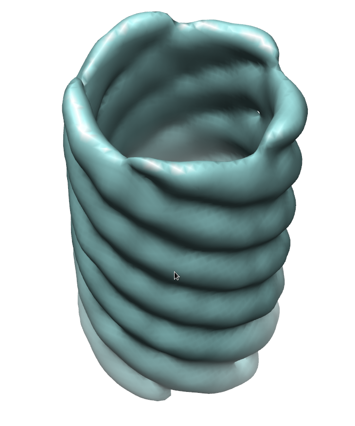
| タイトル | Electron Cryotomography of Measles Virus Reveals how Matrix Protein Coats the Ribonucleocapsid Within Intact Virions. | |||||||||
 マップデータ マップデータ | Measles virus matrix filament | |||||||||
 試料 試料 |
| |||||||||
 キーワード キーワード |  measles (麻疹) / matrix / nucleocapsid (カプシド) / ribonucleocapsid / RNP / MCNC measles (麻疹) / matrix / nucleocapsid (カプシド) / ribonucleocapsid / RNP / MCNC | |||||||||
| 生物種 |   Measles virus (麻疹ウイルス) Measles virus (麻疹ウイルス) | |||||||||
| 手法 | サブトモグラム平均法 / クライオ電子顕微鏡法 / 解像度: 44.0 Å | |||||||||
 データ登録者 データ登録者 | Liljeroos L / Huiskonen JT / Ora A / Susi P / Butcher SJ | |||||||||
 引用 引用 | ジャーナル: Proc Natl Acad Sci U S A / 年: 2011 タイトル: Electron cryotomography of measles virus reveals how matrix protein coats the ribonucleocapsid within intact virions. 著者: Lassi Liljeroos / Juha T Huiskonen / Ari Ora / Petri Susi / Sarah J Butcher /  要旨: Measles virus is a highly infectious, enveloped, pleomorphic virus. We combined electron cryotomography with subvolume averaging and immunosorbent electron microscopy to characterize the 3D ...Measles virus is a highly infectious, enveloped, pleomorphic virus. We combined electron cryotomography with subvolume averaging and immunosorbent electron microscopy to characterize the 3D ultrastructure of the virion. We show that the matrix protein forms helices coating the helical ribonucleocapsid rather than coating the inner leaflet of the membrane, as previously thought. The ribonucleocapsid is folded into tight bundles through matrix-matrix interactions. The implications for virus assembly are that the matrix already tightly interacts with the ribonucleocapsid in the cytoplasm, providing a structural basis for the previously observed regulation of RNA transcription by the matrix protein. Next, the matrix-covered ribonucleocapsids are transported to the plasma membrane, where the matrix interacts with the envelope glycoproteins during budding. These results are relevant to the nucleocapsid organization and budding of other paramyxoviruses, where isolated matrix has been observed to form helices. | |||||||||
| 履歴 |
|
- 構造の表示
構造の表示
| ムービー |
 ムービービューア ムービービューア |
|---|---|
| 構造ビューア | EMマップ: SurfViewMolmilJmol/JSmol |
| 添付画像 |
- ダウンロードとリンク
-EMDBアーカイブ
| マップデータ | emd_1974.map.gz | 1.4 MB | EMDBマップデータ形式 | |
|---|---|---|---|---|
| ヘッダ (付随情報) | emd-1974-v30.xmlemd-1974.xml | 8.2 KB 8.2 KB | 表示 表示 | EMDBヘッダ |
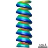
| 画像 |  emd1974.png emd1974.png | 237.4 KB | ||
| アーカイブディレクトリ |  http://ftp.pdbj.org/pub/emdb/structures/EMD-1974ftp://ftp.pdbj.org/pub/emdb/structures/EMD-1974 http://ftp.pdbj.org/pub/emdb/structures/EMD-1974ftp://ftp.pdbj.org/pub/emdb/structures/EMD-1974 | HTTPS FTP |
-関連構造データ
-リンク
| EMDBのページ | EMDB (EBI/PDBe) / EMDataResource |
|---|
-マップ
| ファイル | ダウンロード / ファイル: emd_1974.map.gz / 形式: CCP4 / 大きさ: 1.4 MB / タイプ: IMAGE STORED AS FLOATING POINT NUMBER (4 BYTES) | ||||||||||||||||||||||||||||||||||||||||||||||||||||||||||||||||||||
|---|---|---|---|---|---|---|---|---|---|---|---|---|---|---|---|---|---|---|---|---|---|---|---|---|---|---|---|---|---|---|---|---|---|---|---|---|---|---|---|---|---|---|---|---|---|---|---|---|---|---|---|---|---|---|---|---|---|---|---|---|---|---|---|---|---|---|---|---|---|
| 注釈 | Measles virus matrix filament | ||||||||||||||||||||||||||||||||||||||||||||||||||||||||||||||||||||
| ボクセルのサイズ | X=Y=Z: 7.68 Å | ||||||||||||||||||||||||||||||||||||||||||||||||||||||||||||||||||||
| 密度 |
| ||||||||||||||||||||||||||||||||||||||||||||||||||||||||||||||||||||
| 対称性 | 空間群: 1 | ||||||||||||||||||||||||||||||||||||||||||||||||||||||||||||||||||||
| 詳細 | EMDB XML:
CCP4マップ ヘッダ情報:
| ||||||||||||||||||||||||||||||||||||||||||||||||||||||||||||||||||||
-添付データ
- 試料の構成要素
-全体 : Measles virus matrix-covered ribonucleocapsid, outer helix
| 全体 | 名称: Measles virus matrix-covered ribonucleocapsid, outer helix |
|---|---|
| 要素 |
|
-超分子 #1000: Measles virus matrix-covered ribonucleocapsid, outer helix
| 超分子 | 名称: Measles virus matrix-covered ribonucleocapsid, outer helix タイプ: sample / ID: 1000 / Number unique components: 1 |
|---|
-分子 #1: Matrix Protein
| 分子 | 名称: Matrix Protein / タイプ: protein_or_peptide / ID: 1 / Name.synonym: matrix / 組換発現: No / データベース: NCBI |
|---|---|
| 由来(天然) | 生物種: Measles virus (麻疹ウイルス) |
-実験情報
-構造解析
| 手法 | クライオ電子顕微鏡法 |
|---|---|
 解析 解析 | サブトモグラム平均法 |
| 試料の集合状態 | particle |
-試料調製
| 緩衝液 | pH: 7.4 / 詳細: 20 mM Tris-HCl, 180 mM NaCl, pH 7.4 |
|---|---|
| グリッド | 詳細: C-flat 2/2-2C, holey carbon copper grid |
| 凍結 | 凍結剤: ETHANE / 装置: HOMEMADE PLUNGER / 詳細: Vitrification instrument: Homemade plunger / 手法: Blot for 4 seconds before plunging |
- 電子顕微鏡法
| 顕微鏡 | FEI TECNAI F20 |
|---|---|
| 電子線 | 加速電圧: 200 kV / 電子線源: FIELD EMISSION GUN |
| 電子光学系 | 倍率(補正後): 39400 / 照射モード: FLOOD BEAM / 撮影モード: BRIGHT FIELDBright-field microscopy / Cs: 2.0 mm / 最大 デフォーカス(公称値): 6.0 µm / 最小 デフォーカス(公称値): 6.0 µm / 倍率(公称値): 39400 |
| 試料ステージ | 試料ホルダー: Eucentric / 試料ホルダーモデル: GATAN LIQUID NITROGEN / Tilt series - Axis1 - Min angle: -60 ° / Tilt series - Axis1 - Max angle: 60 ° |
| 撮影 | カテゴリ: CCD フィルム・検出器のモデル: GATAN ULTRASCAN 4000 (4k x 4k) |
| 実験機器 |  モデル: Tecnai F20 / 画像提供: FEI Company |
-画像解析
| 最終 再構成 | 解像度のタイプ: BY AUTHOR / 解像度: 44.0 Å / 解像度の算出法: FSC 0.5 CUT-OFF / ソフトウェア - 名称: IMOD, Bsoft, Jsubtomo |
|---|---|
| 詳細 | Average number of projections used in the 3D reconstructions: 706. |
