色素体 / translation regulator activity / rescue of stalled ribosome / maturation of SSU-rRNA from tricistronic rRNA transcript (SSU-rRNA, 5.8S rRNA, LSU-rRNA) / maturation of SSU-rRNA / protein kinase C binding / modification-dependent protein catabolic process / protein tag activity / ribosomal small subunit assembly / cytosolic small ribosomal subunit ...色素体 / translation regulator activity / rescue of stalled ribosome / maturation of SSU-rRNA from tricistronic rRNA transcript (SSU-rRNA, 5.8S rRNA, LSU-rRNA) / maturation of SSU-rRNA / protein kinase C binding / modification-dependent protein catabolic process / protein tag activity / ribosomal small subunit assembly / cytosolic small ribosomal subunit / ribosome binding / small ribosomal subunit / rRNA binding / protein ubiquitination / リボソーム / structural constituent of ribosome / positive regulation of protein phosphorylation / ribonucleoprotein complex / 翻訳 (生物学) / mRNA binding / ubiquitin protein ligase binding / RNA binding / zinc ion binding / 細胞核 / 細胞質基質 類似検索 - 分子機能
: / Ribosomal protein S12e / Small (40S) ribosomal subunit Asc1/RACK1 / Ribosomal protein S19e, conserved site / S27a-like superfamily / Ribosomal protein S10, eukaryotic/archaeal / Ribosomal protein S25 / 40S ribosomal protein S29/30S ribosomal protein S14 type Z / Ribosomal protein S27a / Ribosomal protein S27a ...: / Ribosomal protein S12e / Small (40S) ribosomal subunit Asc1/RACK1 / Ribosomal protein S19e, conserved site / S27a-like superfamily / Ribosomal protein S10, eukaryotic/archaeal / Ribosomal protein S25 / 40S ribosomal protein S29/30S ribosomal protein S14 type Z / Ribosomal protein S27a / Ribosomal protein S27a / Ribosomal protein S3, eukaryotic/archaeal / S25 ribosomal protein / Ribosomal protein S19A/S15e / Ribosomal protein S12e signature. / Ribosomal protein S17e / Ribosomal protein S17e-like superfamily / Ribosomal protein S27a / Ribosomal protein S19e / Ribosomal_S19e / Ribosomal protein S19e signature. / Ribosomal S17 / Ribosomal protein S19e / 40S Ribosomal protein S10 / Ribosomal protein S28e conserved site / Ribosomal protein S28e / Plectin/S10, N-terminal / Plectin/S10 domain / Ribosomal protein S5/S7, eukaryotic/archaeal / Ribosomal protein S28e / Ribosomal protein S28e signature. / Ribosomal protein L7Ae/L30e/S12e/Gadd45 / Ribosomal protein S14/S29 / Ribosomal protein L7Ae/L30e/S12e/Gadd45 family / 50S ribosomal protein L30e-like / Ubiquitin conserved site / Ubiquitin domain / Ubiquitin domain signature. / Ribosomal protein S3, conserved site / K Homology domain, type 2 / Ribosomal protein S3, C-terminal / Ribosomal protein S3, C-terminal domain superfamily / Ribosomal protein S15/S19, conserved site / KHドメイン / Ribosomal protein S19/S15 / Ribosomal protein S19/S15, superfamily / Ribosomal protein S10 / Ribosomal protein S3, C-terminal domain / Ribosomal protein S3 signature. / Ribosomal protein S7, conserved site / K homology domain superfamily, prokaryotic type / Ribosomal protein S19 / Ribosomal protein S13, conserved site / Ribosomal protein S13 / 30s ribosomal protein S13, C-terminal / Ubiquitin family / Ribosomal protein S14 / Type-2 KH domain profile. / Ribosomal protein S13/S18 / Ribosomal protein S19 signature. / K homology domain-like, alpha/beta / Ribosomal protein S14p/S29e / Ubiquitin homologues / Ribosomal protein S7 signature. / Ribosomal protein S10p/S20e / Ribosomal protein S13-like, H2TH / Ribosomal protein S9, conserved site / Ribosomal protein S10 domain / Ribosomal protein S10 domain superfamily / Ribosomal protein S13 signature. / Ribosomal protein S5/S7 / Ribosomal protein S7 domain / Ribosomal protein S7 domain superfamily / Ribosomal protein S13 family profile. / Ribosomal protein S10p/S20e / Ribosomal protein S9 / ユビキチン様タンパク質 / Ribosomal protein S7p/S5e / Ribosomal protein S9/S16 / Ribosomal protein S9 signature. / Ubiquitin domain profile. / Zinc-binding ribosomal protein / Ribosomal protein S5 domain 2-type fold, subgroup / G-protein beta WD-40 repeat / Ubiquitin-like domain superfamily / Winged helix DNA-binding domain superfamily / WD40 repeat, conserved site / Ribosomal protein S5 domain 2-type fold / Trp-Asp (WD) repeats signature. / WD domain, G-beta repeat / WD40リピート / WD40リピート / Trp-Asp (WD) repeats profile. / Trp-Asp (WD) repeats circular profile. / WD40-repeat-containing domain superfamily / WD40/YVTN repeat-like-containing domain superfamily / Winged helix-like DNA-binding domain superfamily / Nucleic acid-binding, OB-fold 類似検索 - ドメイン・相同性
40S ribosomal protein S17 / 40S ribosomal protein S15 / Small ribosomal subunit protein uS14 / Putative ribosomal protein S18 / Ribosomal protein S20 / Ubiquitin-like domain-containing protein / Plectin/eS10 N-terminal domain-containing protein / Small ribosomal subunit protein uS7c / Uncharacterized protein / 40S ribosomal protein S16 ...40S ribosomal protein S17 / 40S ribosomal protein S15 / Small ribosomal subunit protein uS14 / Putative ribosomal protein S18 / Ribosomal protein S20 / Ubiquitin-like domain-containing protein / Plectin/eS10 N-terminal domain-containing protein / Small ribosomal subunit protein uS7c / Uncharacterized protein / 40S ribosomal protein S16 / 40S ribosomal protein S28 / 40S ribosomal protein S25 / 40S ribosomal protein S12 / 40S ribosomal protein S19 / 30S ribosomal protein S3, chloroplastic 類似検索 - 構成要素
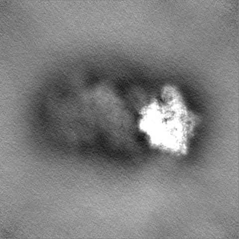
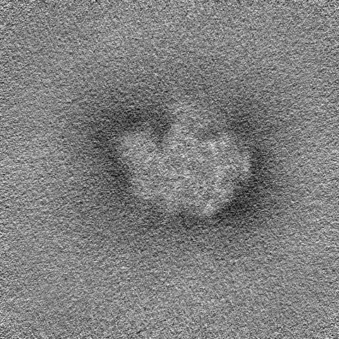
ジャーナル: Int J Mol Sci / 年: 2023 タイトル: High-Resolution Structure and Internal Mobility of a Plant 40S Ribosomal Subunit. 著者: Olesya V Kravchenko / Timur N Baymukhametov / Zhanna A Afonina / Konstantin S Vassilenko / 要旨: Ribosome is a major part of the protein synthesis machinery, and analysis of its structure is of paramount importance. However, the structure of ribosomes from only a limited number of organisms has ...Ribosome is a major part of the protein synthesis machinery, and analysis of its structure is of paramount importance. However, the structure of ribosomes from only a limited number of organisms has been resolved to date; it especially concerns plant ribosomes and ribosomal subunits. Here, we report a high-resolution cryo-electron microscopy reconstruction of the small subunit of the (common wheat) cytoplasmic ribosome. A detailed atomic model was built that includes the majority of the rRNA and some of the protein modifications. The analysis of the obtained data revealed structural peculiarities of the 40S subunit in the monocot plant ribosome. We applied the 3D Flexible Refinement approach to analyze the internal mobility of the 40S subunit and succeeded in decomposing it into four major motions, describing rotations of the head domain and a shift in the massive rRNA expansion segment. It was shown that these motions are almost uncorrelated and that the 40S subunit is flexible enough to spontaneously adopt any conformation it takes as a part of a translating ribosome or ribosomal complex. Here, we introduce the first high-resolution structure of an isolated plant 40S subunit and the first quantitative analysis of the flexibility of small ribosomal subunits, hoping that it will help in studying various aspects of ribosome functioning.
 ムービー
ムービー コントローラー


 データを開く
 基本情報
基本情報
 マップデータ
 試料
試料 キーワード
キーワード wheat (コムギ) /
  機能・相同性情報
 データ登録者
 ロシア, 1件
  引用
引用 構造の表示
構造の表示
 ダウンロードとリンク
 emd_18903.png
 http://ftp.pdbj.org/pub/emdb/structures/EMD-18903


 Z
Z Y
Y X
X


 試料の構成要素
 解析
解析 電子顕微鏡法

