 ムービー
ムービー コントローラー


+ データを開く

- 基本情報
基本情報
| 登録情報 |  | ||||||||||||||||||
|---|---|---|---|---|---|---|---|---|---|---|---|---|---|---|---|---|---|---|---|
| タイトル | wild type neuronal synapse | ||||||||||||||||||
 マップデータ マップデータ | wild type neuronal synapse. non filtered map. | ||||||||||||||||||
 試料 試料 |
| ||||||||||||||||||
 キーワード キーワード |  synapse (シナプス) / neurotransmission / EXOCYTOSIS (エキソサイトーシス) synapse (シナプス) / neurotransmission / EXOCYTOSIS (エキソサイトーシス) | ||||||||||||||||||
| 生物種 |  Mus musculus (ハツカネズミ) Mus musculus (ハツカネズミ) | ||||||||||||||||||
| 手法 | 電子線トモグラフィー法 / クライオ電子顕微鏡法 | ||||||||||||||||||
 データ登録者 データ登録者 | Radecke J / Seeger R / Zuber B / Sorensen JB | ||||||||||||||||||
| 資金援助 |  スイス, スイス,  デンマーク, 5件 デンマーク, 5件
| ||||||||||||||||||
 引用 引用 | ジャーナル: EMBO Rep / 年: 2023 タイトル: Morphofunctional changes at the active zone during synaptic vesicle exocytosis. 著者: Julika Radecke / Raphaela Seeger / Anna Kádková / Ulrike Laugks / Amin Khosrozadeh / Kenneth N Goldie / Vladan Lučić / Jakob B Sørensen / Benoît Zuber /   要旨: Synaptic vesicle (SV) fusion with the plasma membrane (PM) proceeds through intermediate steps that remain poorly resolved. The effect of persistent high or low exocytosis activity on intermediate ...Synaptic vesicle (SV) fusion with the plasma membrane (PM) proceeds through intermediate steps that remain poorly resolved. The effect of persistent high or low exocytosis activity on intermediate steps remains unknown. Using spray-mixing plunge-freezing cryo-electron tomography we observe events following synaptic stimulation at nanometer resolution in near-native samples. Our data suggest that during the stage that immediately follows stimulation, termed early fusion, PM and SV membrane curvature changes to establish a point contact. The next stage-late fusion-shows fusion pore opening and SV collapse. During early fusion, proximal tethered SVs form additional tethers with the PM and increase the inter-SV connector number. In the late-fusion stage, PM-proximal SVs lose their interconnections, allowing them to move toward the PM. Two SNAP-25 mutations, one arresting and one disinhibiting spontaneous release, cause connector loss. The disinhibiting mutation causes loss of membrane-proximal multiple-tethered SVs. Overall, tether formation and connector dissolution are triggered by stimulation and respond to spontaneous fusion rate manipulation. These morphological observations likely correspond to SV transition from one functional pool to another. | ||||||||||||||||||
| 履歴 |
|
- 構造の表示
構造の表示
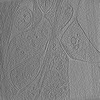
| 添付画像 |
|---|

- ダウンロードとリンク
-EMDBアーカイブ
| マップデータ | emd_18746.map.gz | 310.4 MB |  EMDBマップデータ形式 EMDBマップデータ形式 | |
|---|---|---|---|---|
| ヘッダ (付随情報) | emd-18746-v30.xmlemd-18746.xml | 12.6 KB 12.6 KB | 表示 表示 | EMDBヘッダ |
| 画像 | 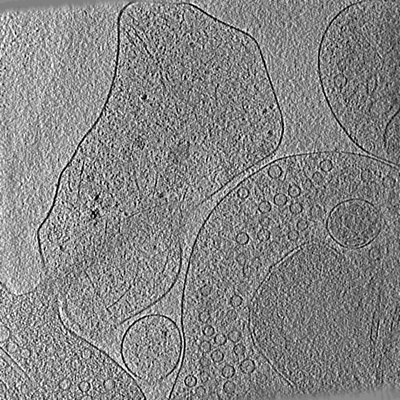 emd_18746.png emd_18746.png | 287 KB | ||
| Filedesc metadata | emd-18746.cif.gz | 4.3 KB | ||
| その他 | emd_18746_additional_1.map.gz | 253.4 MB | ||
| アーカイブディレクトリ |  http://ftp.pdbj.org/pub/emdb/structures/EMD-18746ftp://ftp.pdbj.org/pub/emdb/structures/EMD-18746 http://ftp.pdbj.org/pub/emdb/structures/EMD-18746ftp://ftp.pdbj.org/pub/emdb/structures/EMD-18746 | HTTPS FTP |
-関連構造データ
-リンク
| EMDBのページ | EMDB (EBI/PDBe) / EMDataResource |
|---|
-マップ
| ファイル | ダウンロード / ファイル: emd_18746.map.gz / 形式: CCP4 / 大きさ: 608 MB / タイプ: IMAGE STORED AS SIGNED INTEGER (2 BYTES) | ||||||||||||||||||||
|---|---|---|---|---|---|---|---|---|---|---|---|---|---|---|---|---|---|---|---|---|---|
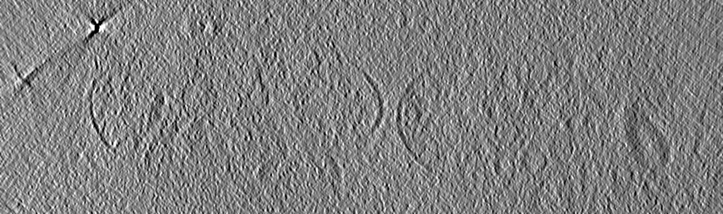
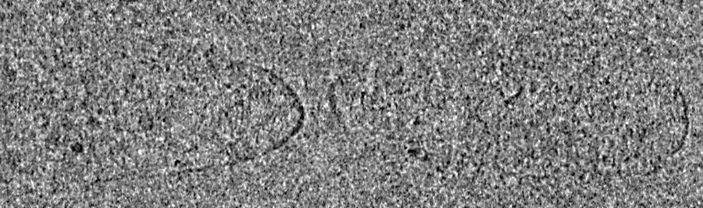
| 注釈 | wild type neuronal synapse. non filtered map. | ||||||||||||||||||||
| ボクセルのサイズ | X=Y=Z: 14.69 Å | ||||||||||||||||||||
| 密度 |
| ||||||||||||||||||||
| 対称性 | 空間群: 1 | ||||||||||||||||||||
| 詳細 | EMDB XML:
|
-添付データ
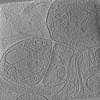
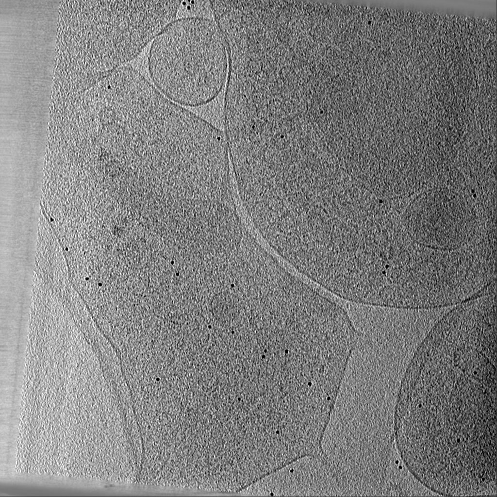
-追加マップ: wild type neuronal synapse. Non-linear anistropic diffusion filtered...
| ファイル | emd_18746_additional_1.map | ||||||||||||
|---|---|---|---|---|---|---|---|---|---|---|---|---|---|
| 注釈 | wild type neuronal synapse. Non-linear anistropic diffusion filtered map | ||||||||||||
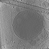
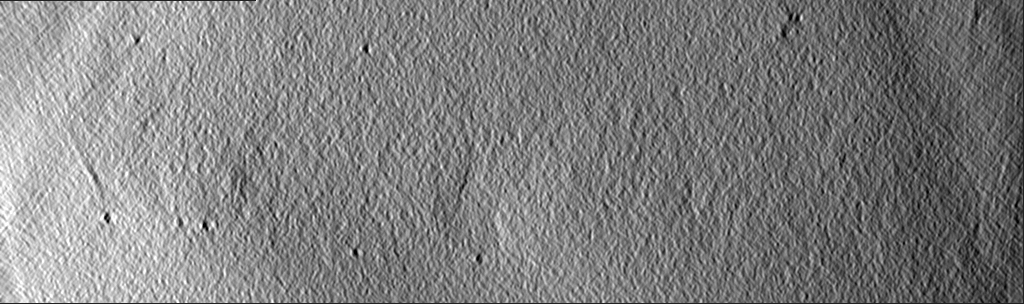
| 投影像・断面図 |
| ||||||||||||
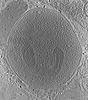
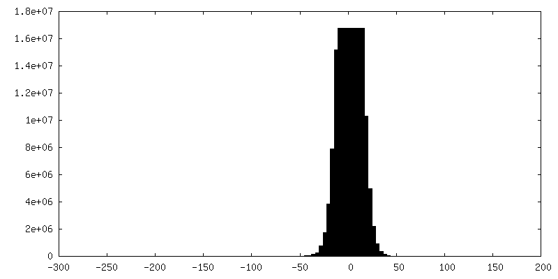
| 密度ヒストグラム |
 Z
Z Y
Y X
X


- 試料の構成要素
-全体 : Synapse between hippocampal neurons isolated from SNAP-25 KO and ...
| 全体 | 名称: Synapse between hippocampal neurons isolated from SNAP-25 KO and transduced with SNAP-25 wild type. |
|---|---|
| 要素 |
|
-超分子 #1: Synapse between hippocampal neurons isolated from SNAP-25 KO and ...
| 超分子 | 名称: Synapse between hippocampal neurons isolated from SNAP-25 KO and transduced with SNAP-25 wild type. タイプ: cell / ID: 1 / 親要素: 0 |
|---|---|
| 由来(天然) | 生物種: Mus musculus (ハツカネズミ) / 株: C57/Bl6 / 器官: Brain / 組織: Hippocampus |
-実験情報
-構造解析
| 手法 | クライオ電子顕微鏡法 |
|---|---|
 解析 解析 | 電子線トモグラフィー法 |
| 試料の集合状態 | cell |
-試料調製
| 緩衝液 | pH: 7.4 詳細: NB medium (Neurobasal with 2% B-27, 1 M HEPES, 0.26% lutamax, 14.3 mM beta-mercaptoethanol, 10000 IU penicillin, 10 mg streptomycin) |
|---|---|
| グリッド | モデル: Quantifoil R2/2 / 材質: GOLD / 支持フィルム - 材質: CARBON / 支持フィルム - トポロジー: HOLEY ARRAY |
| 凍結 | 凍結剤: ETHANE / チャンバー内湿度: 100 % / チャンバー内温度: 277 K / 装置: FEI VITROBOT MARK IV 詳細: After 12 to 14 days of incubation grids with mouse neurons were plunge frozen with a Vitrobot (Thermofisher Scientific, Mark IV) with a blot time of 3 s and a blot force of -10. Wait time and ...詳細: After 12 to 14 days of incubation grids with mouse neurons were plunge frozen with a Vitrobot (Thermofisher Scientific, Mark IV) with a blot time of 3 s and a blot force of -10. Wait time and drain time were not used. Humidity was set to 100% at 4 degrees C. 4 ul undiluted 10 nm BSA gold tracer (Aurion) was added directly onto the grid prior to plunge freezing.. |
| 切片作成 | その他: NO SECTIONING |
| 位置合わせマーカー | Manufacturer: AURION Immuno Gold Reagents & Accessories / 直径: 10 nm |
- 電子顕微鏡法
| 顕微鏡 | FEI TITAN KRIOS |
|---|---|
| 電子線 | 加速電圧: 300 kV / 電子線源: FIELD EMISSION GUN |
| 電子光学系 | 照射モード: FLOOD BEAM / 撮影モード: BRIGHT FIELDBright-field microscopy / 最大 デフォーカス(公称値): 10.0 µm / 最小 デフォーカス(公称値): 6.0 µm |
| 試料ステージ | 試料ホルダーモデル: FEI TITAN KRIOS AUTOGRID HOLDER ホルダー冷却材: NITROGEN |
| 撮影 | フィルム・検出器のモデル: FEI FALCON III (4k x 4k) 検出モード: INTEGRATING / 平均電子線量: 0.76 e/Å2 |
| 実験機器 |  モデル: Titan Krios / 画像提供: FEI Company |
-画像解析
| 最終 再構成 | アルゴリズム: SIMULTANEOUS ITERATIVE (SIRT) / ソフトウェア - 名称: IMOD / 使用した粒子像数: 131 |
|---|
