 ムービー
ムービー コントローラー


+ データを開く

- 基本情報
基本情報
| 登録情報 | データベース: EMDB / ID: EMD-1805 | |||||||||
|---|---|---|---|---|---|---|---|---|---|---|
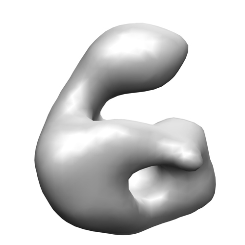
| タイトル | Structure of human complement C8, a precursor to membrane attack. | |||||||||
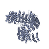
 マップデータ マップデータ | This is the EM reconstruction of human C8 | |||||||||
 試料 試料 |
| |||||||||
 キーワード キーワード | C8 / complement /  membrane attack complex (膜侵襲複合体) / MAC / terminal pathway membrane attack complex (膜侵襲複合体) / MAC / terminal pathway | |||||||||
| 機能・相同性 | Membrane attack complex component/perforin/complement C9 / Thrombospondin type-1 (TSP1) repeat / complement activation, alternative pathway / complement activation, classical pathway / Lipocalin/cytosolic fatty-acid binding domain 機能・相同性情報 機能・相同性情報 | |||||||||
| 生物種 |  Homo sapiens (ヒト) Homo sapiens (ヒト) | |||||||||
| 手法 | 単粒子再構成法 / ネガティブ染色法 / 解像度: 25.0 Å | |||||||||
 データ登録者 データ登録者 | Bubeck D / Roversi P / Donev R / Morgan BP / Llorca O / Lea SM | |||||||||
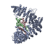
 引用 引用 | ジャーナル: J Mol Biol / 年: 2011 タイトル: Structure of human complement C8, a precursor to membrane attack. 著者: Doryen Bubeck / Pietro Roversi / Rossen Donev / B Paul Morgan / Oscar Llorca / Susan M Lea /  要旨: Complement component C8 plays a pivotal role in the formation of the membrane attack complex (MAC), an important antibacterial immune effector. C8 initiates membrane penetration and coordinates MAC ...Complement component C8 plays a pivotal role in the formation of the membrane attack complex (MAC), an important antibacterial immune effector. C8 initiates membrane penetration and coordinates MAC pore formation. High-resolution structures of C8 subunits have provided some insight into the function of the C8 heterotrimer; however, there is no structural information describing how the intersubunit organization facilitates MAC assembly. We have determined the structure of C8 by electron microscopy and fitted the C8α-MACPF (membrane attack complex/perforin)-C8γ co-crystal structure and a homology model for C8β-MACPF into the density. Here, we demonstrate that both the C8γ protrusion and the C8α-MACPF region that inserts into the membrane upon activation are accessible. | |||||||||
| 履歴 |
|
- 構造の表示
構造の表示
| ムービー |
ムービービューア |
|---|---|
| 構造ビューア | EMマップ: SurfViewMolmilJmol/JSmol |
| 添付画像 |
- ダウンロードとリンク
-EMDBアーカイブ
| マップデータ | emd_1805.map.gz | 126.8 KB | EMDBマップデータ形式 | |
|---|---|---|---|---|
| ヘッダ (付随情報) | emd-1805-v30.xmlemd-1805.xml | 11.3 KB 11.3 KB | 表示 表示 | EMDBヘッダ |
| 画像 |  1805.jpg 1805.jpg | 68.1 KB | ||
| アーカイブディレクトリ |  http://ftp.pdbj.org/pub/emdb/structures/EMD-1805ftp://ftp.pdbj.org/pub/emdb/structures/EMD-1805 http://ftp.pdbj.org/pub/emdb/structures/EMD-1805ftp://ftp.pdbj.org/pub/emdb/structures/EMD-1805 | HTTPS FTP |
-関連構造データ


-リンク
| EMDBのページ | EMDB (EBI/PDBe) / EMDataResource |
|---|
-マップ
| ファイル | ダウンロード / ファイル: emd_1805.map.gz / 形式: CCP4 / 大きさ: 422.9 KB / タイプ: IMAGE STORED AS FLOATING POINT NUMBER (4 BYTES) | ||||||||||||||||||||||||||||||||||||||||||||||||||||||||||||||||||||
|---|---|---|---|---|---|---|---|---|---|---|---|---|---|---|---|---|---|---|---|---|---|---|---|---|---|---|---|---|---|---|---|---|---|---|---|---|---|---|---|---|---|---|---|---|---|---|---|---|---|---|---|---|---|---|---|---|---|---|---|---|---|---|---|---|---|---|---|---|---|
| 注釈 | This is the EM reconstruction of human C8 | ||||||||||||||||||||||||||||||||||||||||||||||||||||||||||||||||||||
| ボクセルのサイズ | X=Y=Z: 4.7 Å | ||||||||||||||||||||||||||||||||||||||||||||||||||||||||||||||||||||
| 密度 |
| ||||||||||||||||||||||||||||||||||||||||||||||||||||||||||||||||||||
| 対称性 | 空間群: 1 | ||||||||||||||||||||||||||||||||||||||||||||||||||||||||||||||||||||
| 詳細 | EMDB XML:
CCP4マップ ヘッダ情報:
| ||||||||||||||||||||||||||||||||||||||||||||||||||||||||||||||||||||
-添付データ
- 試料の構成要素
-全体 : Human complement C8
| 全体 | 名称: Human complement C8 |
|---|---|
| 要素 |
|
-超分子 #1000: Human complement C8
| 超分子 | 名称: Human complement C8 / タイプ: sample / ID: 1000 詳細: The sample was purified by size exclusion chromatography and the purity checked by SDS-PAGE 集合状態: Heterotrimer of C8alpha C8beta and C8gamma / Number unique components: 3 |
|---|---|
| 分子量 | 理論値: 150 KDa |
-分子 #1: Human complement C8 alpha subunit
| 分子 | 名称: Human complement C8 alpha subunit / タイプ: protein_or_peptide / ID: 1 / Name.synonym: C8 alpha / コピー数: 1 / 集合状態: Monomer / 組換発現: No |
|---|---|
| 由来(天然) | 生物種: Homo sapiens (ヒト) / 別称: Human / 組織: Serum / 細胞中の位置: Extracellular |
| 分子量 | 理論値: 65.163 KDa |
| 配列 | GO: complement activation, alternative pathway / InterPro: Thrombospondin type-1 (TSP1) repeat |
-分子 #2: Human complement C8 beta subunit
| 分子 | 名称: Human complement C8 beta subunit / タイプ: protein_or_peptide / ID: 2 / Name.synonym: C8 beta / コピー数: 1 / 集合状態: Monomer / 組換発現: No |
|---|---|
| 由来(天然) | 生物種: Homo sapiens (ヒト) / 別称: Human / 組織: Serum / 細胞中の位置: Extracellular |
| 配列 | GO: complement activation, classical pathway InterPro: Membrane attack complex component/perforin/complement C9 |
-分子 #3: Human complement C8 gamma subunit
| 分子 | 名称: Human complement C8 gamma subunit / タイプ: protein_or_peptide / ID: 3 / Name.synonym: C8 gamma / コピー数: 1 / 集合状態: Monomer / 組換発現: No |
|---|---|
| 由来(天然) | 生物種: Homo sapiens (ヒト) / 別称: Human / 組織: Serum / 細胞中の位置: Extracellular |
| 配列 | GO: complement activation, alternative pathway / InterPro: Lipocalin/cytosolic fatty-acid binding domain |
-実験情報
-構造解析
| 手法 | ネガティブ染色法 |
|---|---|
 解析 解析 | 単粒子再構成法 |
| 試料の集合状態 | particle |
-試料調製
| 濃度 | 0.03 mg/mL |
|---|---|
| 緩衝液 | pH: 7.4 詳細: 100 mM NaCl, 0.15 mM CaCl2, 0.5 mM MgCl2, 5 mM Imidazole pH 7.4. |
| 染色 | タイプ: NEGATIVE / 詳細: 0.75% uranyl formate using the two-drop method |
| グリッド | 詳細: Carbon-coated copper-palladium grid, which was glow-discharged for 10 seconds at 20 mA |
| 凍結 | 凍結剤: NONE / 装置: OTHER |
- 電子顕微鏡法
| 顕微鏡 | FEI TECNAI F30 |
|---|---|
| 電子線 | 加速電圧: 300 kV / 電子線源: OTHER |
| 電子光学系 | 照射モード: OTHER / 撮影モード: OTHER / 倍率(公称値): 59000 |
| 試料ステージ | 試料ホルダー: OTHER / 試料ホルダーモデル: OTHER |
| 撮影 | デジタル化 - スキャナー: ZEISS SCAI / デジタル化 - サンプリング間隔: 7 µm / 平均電子線量: 10 e/Å2 / ビット/ピクセル: 4 |
| 実験機器 |  モデル: Tecnai F30 / 画像提供: FEI Company |
-画像解析
| 最終 2次元分類 | クラス数: 362 |
|---|---|
| 最終 再構成 | 想定した対称性 - 点群: C1 (非対称) / 解像度のタイプ: BY AUTHOR / 解像度: 25.0 Å / 解像度の算出法: FSC 0.5 CUT-OFF / ソフトウェア - 名称: EMAN / 使用した粒子像数: 5167 |
| 詳細 | Micrographs were digitized using a SCAI scanner at a step size of 7 um and binned by a factor of 4 resulting in a pixel size of 4.74 A per pixel |
-原子モデル構築 1
| 初期モデル | PDB ID: Chain - #0 - Chain ID: A / Chain - #1 - Chain ID: C |
|---|---|
| ソフトウェア | 名称: UCSF Chimera |
| 詳細 | PDBEntryID_givenInChain. Protocol: Real space rigid body. Fitted as a rigid body in real space |
| 精密化 | 空間: REAL / プロトコル: RIGID BODY FIT / 当てはまり具合の基準: R-factor |

