 ムービー
ムービー コントローラー


+ データを開く

- 基本情報
基本情報
| 登録情報 |  | ||||||||||||
|---|---|---|---|---|---|---|---|---|---|---|---|---|---|
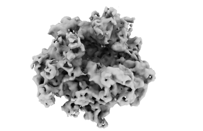
| タイトル | Spraguea lophii ribosome in the closed conformation by cryo sub tomogram averaging | ||||||||||||
 マップデータ マップデータ | B-factor -200 is applied for map sharpening. | ||||||||||||
 試料 試料 |
| ||||||||||||
 キーワード キーワード |  Microsporidia (微胞子虫) / RIBOSOME (リボソーム) Microsporidia (微胞子虫) / RIBOSOME (リボソーム) | ||||||||||||
| 機能・相同性 |  機能・相同性情報 機能・相同性情報translation regulator activity / cytosolic ribosome / ribosome binding / large ribosomal subunit / small ribosomal subunit / 5S rRNA binding / cytosolic large ribosomal subunit / rRNA binding / リボソーム / structural constituent of ribosome ...translation regulator activity / cytosolic ribosome / ribosome binding / large ribosomal subunit / small ribosomal subunit / 5S rRNA binding / cytosolic large ribosomal subunit / rRNA binding / リボソーム / structural constituent of ribosome / 翻訳 (生物学) / ribonucleoprotein complex / RNA binding / zinc ion binding / 生体膜 / metal ion binding / 細胞核 / 細胞質基質 / 細胞質類似検索 - 分子機能 | ||||||||||||
| 生物種 |  Spraguea lophii 42_110 (菌類) Spraguea lophii 42_110 (菌類) | ||||||||||||
| 手法 | サブトモグラム平均法 / クライオ電子顕微鏡法 / 解像度: 10.8 Å | ||||||||||||
 データ登録者 データ登録者 | Gil Diez P / McLaren M / Isupov MN / Daum B / Conners R / Williams B | ||||||||||||
| 資金援助 |  英国, 1件 英国, 1件
| ||||||||||||
 引用 引用 | ジャーナル: Nat Microbiol / 年: 2023 タイトル: CryoEM reveals that ribosomes in microsporidian spores are locked in a dimeric hibernating state. 著者: Mathew McLaren / Rebecca Conners / Michail N Isupov / Patricia Gil-Díez / Lavinia Gambelli / Vicki A M Gold / Andreas Walter / Sean R Connell / Bryony Williams / Bertram Daum /   要旨: Translational control is an essential process for the cell to adapt to varying physiological or environmental conditions. To survive adverse conditions such as low nutrient levels, translation can be ...Translational control is an essential process for the cell to adapt to varying physiological or environmental conditions. To survive adverse conditions such as low nutrient levels, translation can be shut down almost entirely by inhibiting ribosomal function. Here we investigated eukaryotic hibernating ribosomes from the microsporidian parasite Spraguea lophii in situ by a combination of electron cryo-tomography and single-particle electron cryo-microscopy. We show that microsporidian spores contain hibernating ribosomes that are locked in a dimeric (100S) state, which is formed by a unique dimerization mechanism involving the beak region. The ribosomes within the dimer are fully assembled, suggesting that they are ready to be activated once the host cell is invaded. This study provides structural evidence for dimerization acting as a mechanism for ribosomal hibernation in microsporidia, and therefore demonstrates that eukaryotes utilize this mechanism in translational control. | ||||||||||||
| 履歴 |
|
- 構造の表示
構造の表示
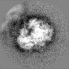
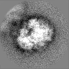
| 添付画像 |
|---|
- ダウンロードとリンク
-EMDBアーカイブ
| マップデータ | emd_17448.map.gz | 656.7 KB | EMDBマップデータ形式 | |
|---|---|---|---|---|
| ヘッダ (付随情報) | emd-17448-v30.xmlemd-17448.xml | 93.3 KB 93.3 KB | 表示 表示 | EMDBヘッダ |
| FSC (解像度算出) | emd_17448_fsc.xml | 3.7 KB | 表示 | FSCデータファイル |
| 画像 |  emd_17448.png emd_17448.png | 50.7 KB | ||
| Filedesc metadata | emd-17448.cif.gz | 17.7 KB | ||
| その他 | emd_17448_half_map_1.map.gzemd_17448_half_map_2.map.gz | 2.8 MB 2.8 MB | ||
| アーカイブディレクトリ |  http://ftp.pdbj.org/pub/emdb/structures/EMD-17448ftp://ftp.pdbj.org/pub/emdb/structures/EMD-17448 http://ftp.pdbj.org/pub/emdb/structures/EMD-17448ftp://ftp.pdbj.org/pub/emdb/structures/EMD-17448 | HTTPS FTP |
-関連構造データ


-リンク
| EMDBのページ | EMDB (EBI/PDBe) / EMDataResource |
|---|---|
| 「今月の分子」の関連する項目 |


-マップ
| ファイル | ダウンロード / ファイル: emd_17448.map.gz / 形式: CCP4 / 大きさ: 3.8 MB / タイプ: IMAGE STORED AS FLOATING POINT NUMBER (4 BYTES) | ||||||||||||||||||||||||||||||||||||
|---|---|---|---|---|---|---|---|---|---|---|---|---|---|---|---|---|---|---|---|---|---|---|---|---|---|---|---|---|---|---|---|---|---|---|---|---|---|
| 注釈 | B-factor -200 is applied for map sharpening. | ||||||||||||||||||||||||||||||||||||
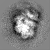
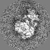
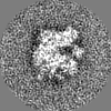
| 投影像・断面図 | 画像のコントロール
画像は Spider により作成 | ||||||||||||||||||||||||||||||||||||
| ボクセルのサイズ | X=Y=Z: 4.53 Å | ||||||||||||||||||||||||||||||||||||
| 密度 |
| ||||||||||||||||||||||||||||||||||||
| 対称性 | 空間群: 1 | ||||||||||||||||||||||||||||||||||||
| 詳細 | EMDB XML:
|
 Z (Sec.)
 Y (Row.)
 X (Col.)


-添付データ
-ハーフマップ: #1
| ファイル | emd_17448_half_map_1.map | ||||||||||||
|---|---|---|---|---|---|---|---|---|---|---|---|---|---|
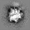
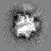
| 投影像・断面図 |
| ||||||||||||
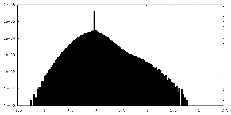
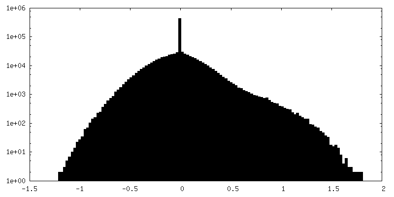
| 密度ヒストグラム |


-ハーフマップ: #2
| ファイル | emd_17448_half_map_2.map | ||||||||||||
|---|---|---|---|---|---|---|---|---|---|---|---|---|---|
| 投影像・断面図 |
| ||||||||||||
| 密度ヒストグラム |


- 試料の構成要素
+全体 : Ribosome
+超分子 #1: Ribosome
+分子 #1: RNA 28S
+分子 #2: RNA 5S
+分子 #42: RNA 16S
+分子 #3: 60S ribosomal protein L8
+分子 #4: uL15
+分子 #5: 60S ribosomal protein L3
+分子 #6: 60S ribosomal protein L4
+分子 #7: 60S ribosomal protein L3
+分子 #8: 60S ribosomal protein L5
+分子 #9: 60S ribosomal protein L31
+分子 #10: 60S ribosomal protein L6
+分子 #11: 60S ribosomal protein L32
+分子 #12: 60S ribosomal protein L7
+分子 #13: 60S ribosomal protein L35a
+分子 #14: 60S ribosomal protein L8
+分子 #15: Ribosomal protein L34
+分子 #16: 60S ribosomal protein L9
+分子 #17: Ribosomal L29 protein (Fragment),Ribosomal L29 protein (Fragment)...
+分子 #18: S60 ribosomal protein L10
+分子 #19: 60S ribosomal protein L36
+分子 #20: 60S ribosomal protein L11
+分子 #21: eL37
+分子 #22: 60S ribosomal protein L13
+分子 #23: 60S ribosomal protein L39
+分子 #24: Transposase
+分子 #25: Ubiquitin
+分子 #26: Ribosomal protein L15
+分子 #27: Ribosomal protein L13A
+分子 #28: 60S ribosomal protein L44
+分子 #29: 60S ribosomal protein L17
+分子 #30: 60S ribosomal protein L37a
+分子 #31: 60S ribosomal protein L18
+分子 #32: 60S ribosomal protein L19
+分子 #33: 60S ribosomal protein L20
+分子 #34: 60s ribosomal protein L21
+分子 #35: 60S ribosomal protein L22
+分子 #36: Ribosomal protein L23
+分子 #37: Ribosomal protein L24E
+分子 #38: 60S ribosomal protein L23a
+分子 #39: 60S ribosomal protein L26
+分子 #40: 60S ribosomal protein L27
+分子 #41: DNL-type domain-containing protein
+分子 #43: 40S ribosomal protein S0
+分子 #44: 40S ribosomal protein S26
+分子 #45: eS1
+分子 #46: eS27
+分子 #47: 40S ribosomal protein S2
+分子 #48: eS28
+分子 #49: 40S ribosomal protein S3
+分子 #50: 40S ribosomal protein S29
+分子 #51: 40S ribosomal protein S4
+分子 #52: eS30
+分子 #53: 40S ribosomal protein S5
+分子 #54: Ubiquitin/40s ribosomal protein S27a fusion
+分子 #55: 40S ribosomal protein S6
+分子 #56: Guanine nucleotide binding protein beta subunit
+分子 #57: 40S ribosomal protein S7
+分子 #58: 40S ribosomal protein S8
+分子 #59: 40S ribosomal protein S9
+分子 #60: 40S ribosomal protein S10
+分子 #61: 40S ribosomal protein S11
+分子 #62: 40S ribosomal protein S12
+分子 #63: 40S ribosomal protein S13
+分子 #64: 40S ribosomal protein S14
+分子 #65: Ribosomal protein S19
+分子 #66: 40S ribosomal protein S16
+分子 #67: eS17
+分子 #68: 40S ribosomal protein S18
+分子 #69: 40S Ribosomal protein S19
+分子 #70: 40S ribosomal protein S20
+分子 #71: Ribosomal protein S21E
+分子 #72: 40S ribosomal protein S15A
+分子 #73: uS12
+分子 #74: 40s ribosomal protein s24
+分子 #75: 40S ribosomal protein S25
+分子 #76: ZINC ION
-実験情報
-構造解析
| 手法 | クライオ電子顕微鏡法 |
|---|---|
 解析 解析 | サブトモグラム平均法 |
| 試料の集合状態 | particle |
-試料調製
| 緩衝液 | pH: 7.5 |
|---|---|
| グリッド | モデル: Quantifoil / 材質: COPPER / メッシュ: 300 / 支持フィルム - 材質: CARBON / 支持フィルム - トポロジー: CONTINUOUS / 支持フィルム - Film thickness: 2 / 詳細: 20 mA, Carbon coated grid |
| 凍結 | 凍結剤: ETHANE / チャンバー内湿度: 100 % / チャンバー内温度: 288.15 K / 装置: FEI VITROBOT MARK IV / 詳細: blot force -1 and blot time 4 s. |
- 電子顕微鏡法
| 顕微鏡 | FEI TITAN KRIOS |
|---|---|
| 電子線 | 加速電圧: 300 kV / 電子線源: FIELD EMISSION GUN |
| 電子光学系 | 照射モード: FLOOD BEAM / 撮影モード: BRIGHT FIELDBright-field microscopy / Cs: 2.7 mm / 最大 デフォーカス(公称値): 6.0 µm / 最小 デフォーカス(公称値): 2.5 µm |
| 試料ステージ | 試料ホルダーモデル: FEI TITAN KRIOS AUTOGRID HOLDER ホルダー冷却材: NITROGEN |
| 撮影 | #0 - Image recording ID: 1 #0 - フィルム・検出器のモデル: GATAN K2 SUMMIT (4k x 4k) #0 - 検出モード: COUNTING / #0 - 平均電子線量: 1.98 e/Å2 / #1 - Image recording ID: 2 #1 - フィルム・検出器のモデル: GATAN K3 BIOQUANTUM (6k x 4k) #1 - 平均電子線量: 1.98 e/Å2 |
| 実験機器 |  モデル: Titan Krios / 画像提供: FEI Company |
-画像解析
| 抽出 | トモグラム数: 20 / 使用した粒子像数: 6505 ソフトウェア: (名称: IMOD (ver. 4.12.10), Warp (ver. 1.1.0)) |
|---|---|
| 最終 角度割当 | タイプ: MAXIMUM LIKELIHOOD / ソフトウェア - 名称: RELION (ver. 3.1) |
| 最終 再構成 | 解像度のタイプ: BY AUTHOR / 解像度: 10.8 Å / 解像度の算出法: FSC 0.143 CUT-OFF ソフトウェア: (名称: RELION (ver. 3.1), Warp (ver. 1.1.0)) 使用したサブトモグラム数: 1344 |
| Image recording ID | 2 |
| FSC曲線 (解像度の算出) | 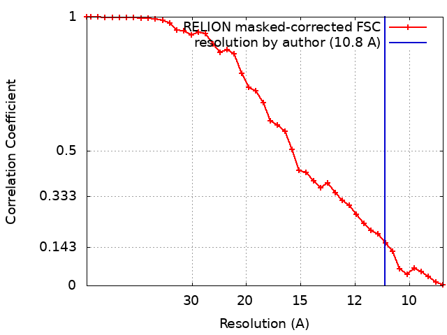 |
-原子モデル構築 1
| 精密化 | 空間: REAL / プロトコル: RIGID BODY FIT |
|---|---|
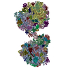
| 得られたモデル | 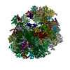 PDB-8p5d: |
