 ムービー
ムービー コントローラー


+ データを開く

- 基本情報
基本情報
| 登録情報 | データベース: EMDB / ID: EMD-1732 | |||||||||
|---|---|---|---|---|---|---|---|---|---|---|
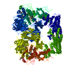
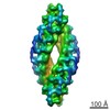
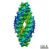
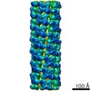
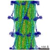
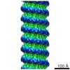
| タイトル | Three-dimensional structure of Tripeptidyl peptidase II from Drosophila melanogaster - a spindle shaped homo-40mer | |||||||||
 マップデータ マップデータ | EM density map of TPPII from Drosophila melanogaster | |||||||||
 試料 試料 |
| |||||||||
 キーワード キーワード |  Protease (プロテアーゼ) / subtilase (スブチラーゼ) Protease (プロテアーゼ) / subtilase (スブチラーゼ) | |||||||||
| 生物種 |  Drosophila melanogaster (キイロショウジョウバエ) Drosophila melanogaster (キイロショウジョウバエ) | |||||||||
| 手法 | 単粒子再構成法 / クライオ電子顕微鏡法 / 解像度: 14.0 Å | |||||||||
 データ登録者 データ登録者 | Chuang CK / Rockel B / Seyit G / Walian PJ / Schoenegge A-M / Peters J / Zwart PH / Baumeister W / Jap BK | |||||||||
 引用 引用 | ジャーナル: Nat Struct Mol Biol / 年: 2010 タイトル: Hybrid molecular structure of the giant protease tripeptidyl peptidase II. 著者: Crystal K Chuang / Beate Rockel / Gönül Seyit / Peter J Walian / Anne-Marie Schönegge / Jürgen Peters / Petrus H Zwart / Wolfgang Baumeister / Bing K Jap /  要旨: Tripeptidyl peptidase II (TPP II) is the largest known eukaryotic protease (6 MDa). It is believed to act downstream of the 26S proteasome, cleaving tripeptides from the N termini of longer peptides, ...Tripeptidyl peptidase II (TPP II) is the largest known eukaryotic protease (6 MDa). It is believed to act downstream of the 26S proteasome, cleaving tripeptides from the N termini of longer peptides, and it is implicated in numerous cellular processes. Here we report the structure of Drosophila TPP II determined by a hybrid approach. We solved the structure of the dimer by X-ray crystallography and docked it into the three-dimensional map of the holocomplex, which we obtained by single-particle cryo-electron microscopy. The resulting structure reveals the compartmentalization of the active sites inside a system of chambers and suggests the existence of a molecular ruler determining the size of the cleavage products. Furthermore, the structure suggests a model for activation of TPP II involving the relocation of a flexible loop and a repositioning of the active-site serine, coupling it to holocomplex assembly and active-site sequestration. | |||||||||
| 履歴 |
|
- 構造の表示
構造の表示
| ムービー |
 ムービービューア ムービービューア |
|---|---|
| 構造ビューア | EMマップ: SurfViewMolmilJmol/JSmol |
| 添付画像 |

- ダウンロードとリンク
-EMDBアーカイブ
| マップデータ | emd_1732.map.gz | 113.3 MB | EMDBマップデータ形式 | |
|---|---|---|---|---|
| ヘッダ (付随情報) | emd-1732-v30.xmlemd-1732.xml | 8.4 KB 8.4 KB | 表示 表示 | EMDBヘッダ |
| 画像 | 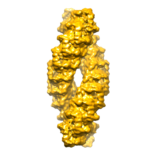 emd1732.png emd1732.png | 136.8 KB | ||
| アーカイブディレクトリ |  http://ftp.pdbj.org/pub/emdb/structures/EMD-1732ftp://ftp.pdbj.org/pub/emdb/structures/EMD-1732 http://ftp.pdbj.org/pub/emdb/structures/EMD-1732ftp://ftp.pdbj.org/pub/emdb/structures/EMD-1732 | HTTPS FTP |
-関連構造データ
-リンク
| EMDBのページ | EMDB (EBI/PDBe) / EMDataResource |
|---|
-マップ
| ファイル | ダウンロード / ファイル: emd_1732.map.gz / 形式: CCP4 / 大きさ: 122.1 MB / タイプ: IMAGE STORED AS FLOATING POINT NUMBER (4 BYTES) | ||||||||||||||||||||||||||||||||||||||||||||||||||||||||||||||||||||
|---|---|---|---|---|---|---|---|---|---|---|---|---|---|---|---|---|---|---|---|---|---|---|---|---|---|---|---|---|---|---|---|---|---|---|---|---|---|---|---|---|---|---|---|---|---|---|---|---|---|---|---|---|---|---|---|---|---|---|---|---|---|---|---|---|---|---|---|---|---|
| 注釈 | EM density map of TPPII from Drosophila melanogaster | ||||||||||||||||||||||||||||||||||||||||||||||||||||||||||||||||||||
| 投影像・断面図 | 画像のコントロール
画像は Spider により作成 | ||||||||||||||||||||||||||||||||||||||||||||||||||||||||||||||||||||
| ボクセルのサイズ | X=Y=Z: 2.16 Å | ||||||||||||||||||||||||||||||||||||||||||||||||||||||||||||||||||||
| 密度 |
| ||||||||||||||||||||||||||||||||||||||||||||||||||||||||||||||||||||
| 対称性 | 空間群: 1 | ||||||||||||||||||||||||||||||||||||||||||||||||||||||||||||||||||||
| 詳細 | EMDB XML:
CCP4マップ ヘッダ情報:
| ||||||||||||||||||||||||||||||||||||||||||||||||||||||||||||||||||||
 Z (Sec.)
 Y (Row.)
 X (Col.)


-添付データ
- 試料の構成要素
-全体 : Tripeptidyl peptidase II complex of Drosophila melanogaster
| 全体 | 名称: Tripeptidyl peptidase II complex of Drosophila melanogaster |
|---|---|
| 要素 |
|
-超分子 #1000: Tripeptidyl peptidase II complex of Drosophila melanogaster
| 超分子 | 名称: Tripeptidyl peptidase II complex of Drosophila melanogaster タイプ: sample / ID: 1000 / 集合状態: 40-mer / Number unique components: 1 |
|---|---|
| 分子量 | 理論値: 6 MDa |
-分子 #1: Tripeptidyl peptidase II
| 分子 | 名称: Tripeptidyl peptidase II / タイプ: protein_or_peptide / ID: 1 / Name.synonym: TPPII / コピー数: 40 / 集合状態: 40-mer / 組換発現: Yes |
|---|---|
| 由来(天然) | 生物種: Drosophila melanogaster (キイロショウジョウバエ) 別称: Fruit Fly / 細胞中の位置: Cytoplasmic |
| 分子量 | 理論値: 150 KDa |
| 組換発現 | 生物種:  Escherichia coli (大腸菌) Escherichia coli (大腸菌) |
-実験情報
-構造解析
| 手法 | クライオ電子顕微鏡法 |
|---|---|
 解析 解析 | 単粒子再構成法 |
| 試料の集合状態 | particle |
-試料調製
| 緩衝液 | pH: 7.8 / 詳細: 40 mM KPO4, pH 7.8, 2 mM DTT, 5% glycerol |
|---|---|
| グリッド | 詳細: Holey carbon 200 mesh copper grids covered with thin carbon film |
| 凍結 | 凍結剤: ETHANE / 装置: OTHER 手法: Sample was applied on grid, blotted briefly and washed twice with buffer (40 mM ammonium sulfate, pH 7.5) |
- 電子顕微鏡法
| 顕微鏡 | FEI/PHILIPS CM200FEG |
|---|---|
| 電子線 | 加速電圧: 160 kV / 電子線源: FIELD EMISSION GUN |
| 電子光学系 | 倍率(補正後): 69500 / 照射モード: FLOOD BEAM / 撮影モード: BRIGHT FIELDBright-field microscopy / 最大 デフォーカス(公称値): 3.0 µm / 最小 デフォーカス(公称値): 0.7 µm |
| 試料ステージ | 試料ホルダー: Side-entry / 試料ホルダーモデル: GATAN LIQUID NITROGEN |
| 撮影 | カテゴリ: CCD フィルム・検出器のモデル: GENERIC TVIPS (4k x 4k) 平均電子線量: 15 e/Å2 |
-画像解析
| CTF補正 | 詳細: Each CCD frame |
|---|---|
| 最終 再構成 | 想定した対称性 - 点群: D2 (2回x2回 2面回転対称) 解像度のタイプ: BY AUTHOR / 解像度: 14.0 Å / 解像度の算出法: FSC 0.5 CUT-OFF / ソフトウェア - 名称: EMAN / 使用した粒子像数: 37195 |
