 ムービー
ムービー コントローラー


+ データを開く

- 基本情報
基本情報
| 登録情報 | データベース: EMDB / ID: EMD-1581 | |||||||||
|---|---|---|---|---|---|---|---|---|---|---|
| タイトル | Structure and functional role of dynein's microtubule-binding domain | |||||||||
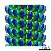
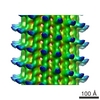
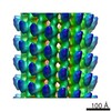
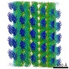
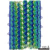
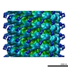
 マップデータ マップデータ | This is a 3-D map of the SRS-MTBD construct | |||||||||
 試料 試料 |
| |||||||||
 キーワード キーワード | Assembly 1 is a helical 15 protofilaments / component name / tubulin. Assembly 2 is a monomer of the SRS-MTBD construct | |||||||||
| 生物種 |   Mus musculus (ハツカネズミ) Mus musculus (ハツカネズミ) | |||||||||
| 手法 | らせん対称体再構成法 / クライオ電子顕微鏡法 / 解像度: 35.0 Å | |||||||||
 データ登録者 データ登録者 | Carter AP / Garbarino JE / Wilson-Kubalek EM / Shipley WE / Cho C / Milligan RA / Vale RD / Gibbons IR | |||||||||
 引用 引用 | ジャーナル: Science / 年: 2008 タイトル: Structure and functional role of dynein's microtubule-binding domain. 著者: Andrew P Carter / Joan E Garbarino / Elizabeth M Wilson-Kubalek / Wesley E Shipley / Carol Cho / Ronald A Milligan / Ronald D Vale / I R Gibbons /  要旨: Dynein motors move various cargos along microtubules within the cytoplasm and power the beating of cilia and flagella. An unusual feature of dynein is that its microtubule-binding domain (MTBD) is ...Dynein motors move various cargos along microtubules within the cytoplasm and power the beating of cilia and flagella. An unusual feature of dynein is that its microtubule-binding domain (MTBD) is separated from its ring-shaped AAA+ adenosine triphosphatase (ATPase) domain by a 15-nanometer coiled-coil stalk. We report the crystal structure of the mouse cytoplasmic dynein MTBD and a portion of the coiled coil, which supports a mechanism by which the ATPase domain and MTBD may communicate through a shift in the heptad registry of the coiled coil. Surprisingly, functional data suggest that the MTBD, and not the ATPase domain, is the main determinant of the direction of dynein motility. | |||||||||
| 履歴 |
|
- 構造の表示
構造の表示
| ムービー |
 ムービービューア ムービービューア |
|---|---|
| 構造ビューア | EMマップ: SurfViewMolmilJmol/JSmol |
| 添付画像 |

- ダウンロードとリンク
-EMDBアーカイブ
| マップデータ | emd_1581.map.gz | 1.3 MB | EMDBマップデータ形式 | |
|---|---|---|---|---|
| ヘッダ (付随情報) | emd-1581-v30.xmlemd-1581.xml | 9.7 KB 9.7 KB | 表示 表示 | EMDBヘッダ |
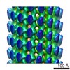
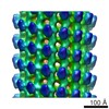
| 画像 | 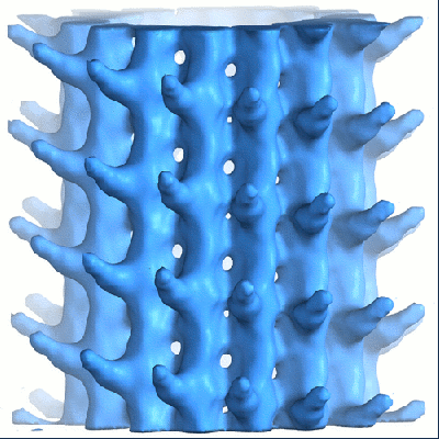 1581.gif 1581.gif | 103.1 KB | ||
| アーカイブディレクトリ |  http://ftp.pdbj.org/pub/emdb/structures/EMD-1581ftp://ftp.pdbj.org/pub/emdb/structures/EMD-1581 http://ftp.pdbj.org/pub/emdb/structures/EMD-1581ftp://ftp.pdbj.org/pub/emdb/structures/EMD-1581 | HTTPS FTP |
-関連構造データ
-リンク
| EMDBのページ | EMDB (EBI/PDBe) / EMDataResource |
|---|
-マップ
| ファイル | ダウンロード / ファイル: emd_1581.map.gz / 形式: CCP4 / 大きさ: 7.8 MB / タイプ: IMAGE STORED AS FLOATING POINT NUMBER (4 BYTES) | ||||||||||||||||||||||||||||||||||||||||||||||||||||||||||||||||||||
|---|---|---|---|---|---|---|---|---|---|---|---|---|---|---|---|---|---|---|---|---|---|---|---|---|---|---|---|---|---|---|---|---|---|---|---|---|---|---|---|---|---|---|---|---|---|---|---|---|---|---|---|---|---|---|---|---|---|---|---|---|---|---|---|---|---|---|---|---|---|
| 注釈 | This is a 3-D map of the SRS-MTBD construct | ||||||||||||||||||||||||||||||||||||||||||||||||||||||||||||||||||||
| ボクセルのサイズ | X=Y=Z: 3.5 Å | ||||||||||||||||||||||||||||||||||||||||||||||||||||||||||||||||||||
| 密度 |
| ||||||||||||||||||||||||||||||||||||||||||||||||||||||||||||||||||||
| 対称性 | 空間群: 1 | ||||||||||||||||||||||||||||||||||||||||||||||||||||||||||||||||||||
| 詳細 | EMDB XML:
CCP4マップ ヘッダ情報:
| ||||||||||||||||||||||||||||||||||||||||||||||||||||||||||||||||||||
-添付データ
- 試料の構成要素
-全体 : Synthetic construct of dynein microtubule-binding domain (85-82) ...
| 全体 | 名称: Synthetic construct of dynein microtubule-binding domain (85-82) fused to seryl-tRNA synthase-monomer. Abbreviated name is SRS-MTBD-85-82 |
|---|---|
| 要素 |
|
-超分子 #1000: Synthetic construct of dynein microtubule-binding domain (85-82) ...
| 超分子 | 名称: Synthetic construct of dynein microtubule-binding domain (85-82) fused to seryl-tRNA synthase-monomer. Abbreviated name is SRS-MTBD-85-82 タイプ: sample / ID: 1000 詳細: The SRS-MTBD-85-82 construct has a 12 heptad long stalk, only the first 3 heptad repeats were visible in this map. No density was observed for the SRS. 集合状態: SRS-MTBD-85-82 monomers bound to 15 protofilaments helical microtubules Number unique components: 2 |
|---|
-分子 #1: microtubule
| 分子 | 名称: microtubule / タイプ: protein_or_peptide / ID: 1 / Name.synonym: microtubule / 集合状態: monomer / 組換発現: Yes |
|---|---|
| 由来(天然) | 生物種: Mus musculus (ハツカネズミ) / 別称: House Mouse |
| 組換発現 | 生物種:  Escherichia coli (大腸菌) Escherichia coli (大腸菌) |
-実験情報
-構造解析
| 手法 | クライオ電子顕微鏡法 |
|---|---|
 解析 解析 | らせん対称体再構成法 |
| 試料の集合状態 | filament |
-試料調製
| 凍結 | 凍結剤: ETHANE / チャンバー内湿度: 90 % / チャンバー内温度: 4 K / 装置: OTHER / 詳細: Vitrification instrument: Vitrobot / 手法: 1.5 sec blot |
|---|
- 電子顕微鏡法
| 顕微鏡 | FEI TECNAI F20 |
|---|---|
| 電子線 | 加速電圧: 120 kV / 電子線源: FIELD EMISSION GUN |
| 電子光学系 | 照射モード: FLOOD BEAM / 撮影モード: BRIGHT FIELDBright-field microscopy / Cs: 2 mm / 倍率(公称値): 29000 |
| 特殊光学系 | エネルギーフィルター - 名称: Field Emission Gun |
| 試料ステージ | 試料ホルダー: eucentric / 試料ホルダーモデル: GATAN LIQUID NITROGEN |
| 撮影 | カテゴリ: CCD フィルム・検出器のモデル: GATAN ULTRASCAN 4000 (4k x 4k) デジタル化 - サンプリング間隔: 3.5 µm / 実像数: 10 / 平均電子線量: 10 e/Å2 / ビット/ピクセル: 8 |
| 実験機器 |  モデル: Tecnai F20 / 画像提供: FEI Company |
-画像解析
| CTF補正 | 詳細: each image |
|---|---|
| 最終 再構成 | アルゴリズム: OTHER / 解像度のタイプ: BY AUTHOR / 解像度: 35.0 Å / 解像度の算出法: OTHER / ソフトウェア - 名称: phoelix |
-原子モデル構築 1
| 初期モデル | PDB ID: Chain - Chain ID: A |
|---|---|
| 詳細 | PDBEntryID_givenInChain. Protocol: Rigid body. The crystal structure was manually docked into the EM density using the chimera software package. |
| 精密化 | 空間: REAL / プロトコル: RIGID BODY FIT |
