 ムービー
ムービー コントローラー


+ データを開く

- 基本情報
基本情報
| 登録情報 |  | |||||||||
|---|---|---|---|---|---|---|---|---|---|---|
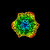
| タイトル | Structure of human Apoferritin obtained from ssDNA coated grid | |||||||||
 マップデータ マップデータ | Final Map | |||||||||
 試料 試料 |
| |||||||||
| 機能・相同性 |  機能・相同性情報 機能・相同性情報iron ion sequestering activity / : /  オートファゴソーム / Scavenging by Class A Receptors / Golgi Associated Vesicle Biogenesis / ferroxidase / intracellular sequestering of iron ion / ferroxidase activity / negative regulation of fibroblast proliferation / ferric iron binding ...iron ion sequestering activity / : / オートファゴソーム / Scavenging by Class A Receptors / Golgi Associated Vesicle Biogenesis / ferroxidase / intracellular sequestering of iron ion / ferroxidase activity / negative regulation of fibroblast proliferation / ferric iron binding / ferrous iron binding / Iron uptake and transport / tertiary granule lumen / iron ion transport / intracellular iron ion homeostasis / ficolin-1-rich granule lumen / 免疫応答 / iron ion binding / negative regulation of cell population proliferation / Neutrophil degranulation / extracellular exosome / extracellular region / identical protein binding / 細胞核 / 細胞質基質 / 細胞質 オートファゴソーム / Scavenging by Class A Receptors / Golgi Associated Vesicle Biogenesis / ferroxidase / intracellular sequestering of iron ion / ferroxidase activity / negative regulation of fibroblast proliferation / ferric iron binding ...iron ion sequestering activity / : / オートファゴソーム / Scavenging by Class A Receptors / Golgi Associated Vesicle Biogenesis / ferroxidase / intracellular sequestering of iron ion / ferroxidase activity / negative regulation of fibroblast proliferation / ferric iron binding / ferrous iron binding / Iron uptake and transport / tertiary granule lumen / iron ion transport / intracellular iron ion homeostasis / ficolin-1-rich granule lumen / 免疫応答 / iron ion binding / negative regulation of cell population proliferation / Neutrophil degranulation / extracellular exosome / extracellular region / identical protein binding / 細胞核 / 細胞質基質 / 細胞質類似検索 - 分子機能 | |||||||||
| 生物種 |  Homo sapiens (ヒト) Homo sapiens (ヒト) | |||||||||
| 手法 | 単粒子再構成法 / クライオ電子顕微鏡法 / 解像度: 1.77 Å | |||||||||
 データ登録者 データ登録者 | Hrebik D / Plevka P | |||||||||
| 資金援助 |  チェコ, 1件 チェコ, 1件
| |||||||||
 引用 引用 | ジャーナル: Acta Crystallogr D Struct Biol / 年: 2022 タイトル: Polyelectrolyte coating of cryo-EM grids improves lateral distribution and prevents aggregation of macromolecules. 著者: Dominik Hrebík / Mária Gondová / Lucie Valentová / Tibor Füzik / Antonín Přidal / Jiří Nováček / Pavel Plevka / 要旨: Cryo-electron microscopy (cryo-EM) is one of the primary methods used to determine the structures of macromolecules and their complexes. With the increased availability of cryo-electron microscopes, ...Cryo-electron microscopy (cryo-EM) is one of the primary methods used to determine the structures of macromolecules and their complexes. With the increased availability of cryo-electron microscopes, the preparation of high-quality samples has become a bottleneck in the cryo-EM structure-determination pipeline. Macromolecules can be damaged during the purification or preparation of vitrified samples for cryo-EM, making them prone to binding to the grid support, to aggregation or to the adoption of preferential orientations at the air-water interface. Here, it is shown that coating cryo-EM grids with a negatively charged polyelectrolyte, such as single-stranded DNA, before applying the sample reduces the aggregation of macromolecules and improves their distribution. The single-stranded DNA-coated grids enabled the determination of high-resolution structures from samples that aggregated on conventional grids. The polyelectrolyte coating reduces the diffusion of macromolecules and thus may limit the negative effects of the contact of macromolecules with the grid support and blotting paper, as well as of the shear forces on macromolecules during grid blotting. Coating grids with polyelectrolytes can readily be employed in any laboratory dealing with cryo-EM sample preparation, since it is fast, simple, inexpensive and does not require specialized equipment. | |||||||||
| 履歴 |
|
- 構造の表示
構造の表示
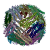
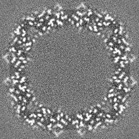
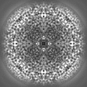
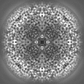
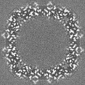
| 添付画像 |
|---|

- ダウンロードとリンク
-EMDBアーカイブ
| マップデータ | emd_14705.map.gz | 71.8 MB | EMDBマップデータ形式 | |
|---|---|---|---|---|
| ヘッダ (付随情報) | emd-14705-v30.xmlemd-14705.xml | 19.9 KB 19.9 KB | 表示 表示 | EMDBヘッダ |
| FSC (解像度算出) | emd_14705_fsc.xml | 18.1 KB | 表示 | FSCデータファイル |
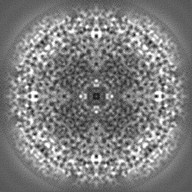
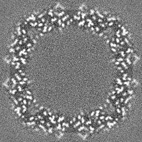
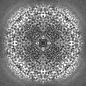
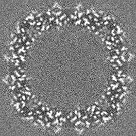
| 画像 | 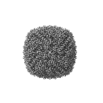 emd_14705.png emd_14705.png | 82.6 KB | ||
| その他 | emd_14705_half_map_1.map.gzemd_14705_half_map_2.map.gz | 77.9 MB 77.9 MB | ||
| アーカイブディレクトリ |  http://ftp.pdbj.org/pub/emdb/structures/EMD-14705ftp://ftp.pdbj.org/pub/emdb/structures/EMD-14705 http://ftp.pdbj.org/pub/emdb/structures/EMD-14705ftp://ftp.pdbj.org/pub/emdb/structures/EMD-14705 | HTTPS FTP |
-関連構造データ
-リンク
| EMDBのページ | EMDB (EBI/PDBe) / EMDataResource |
|---|---|
| 「今月の分子」の関連する項目 |


-マップ
| ファイル | ダウンロード / ファイル: emd_14705.map.gz / 形式: CCP4 / 大きさ: 83.7 MB / タイプ: IMAGE STORED AS FLOATING POINT NUMBER (4 BYTES) | ||||||||||||||||||||
|---|---|---|---|---|---|---|---|---|---|---|---|---|---|---|---|---|---|---|---|---|---|
| 注釈 | Final Map | ||||||||||||||||||||
| ボクセルのサイズ | X=Y=Z: 0.4525 Å | ||||||||||||||||||||
| 密度 |
| ||||||||||||||||||||
| 対称性 | 空間群: 1 | ||||||||||||||||||||
| 詳細 | EMDB XML:
|
-添付データ
-ハーフマップ: Half map 1
| ファイル | emd_14705_half_map_1.map | ||||||||||||
|---|---|---|---|---|---|---|---|---|---|---|---|---|---|
| 注釈 | Half map 1 | ||||||||||||
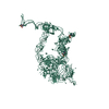
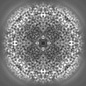
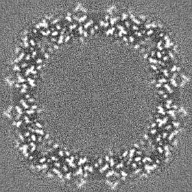
| 投影像・断面図 |
| ||||||||||||
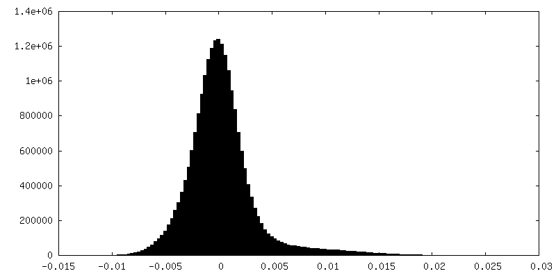
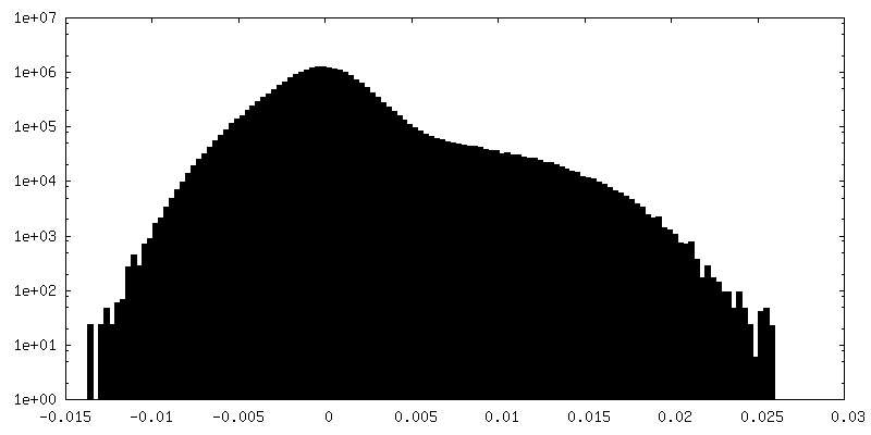
| 密度ヒストグラム |
 Z
Z Y
Y X
X

-ハーフマップ: Half map 2
| ファイル | emd_14705_half_map_2.map | ||||||||||||
|---|---|---|---|---|---|---|---|---|---|---|---|---|---|
| 注釈 | Half map 2 | ||||||||||||
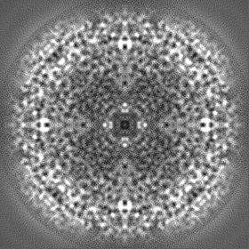
| 投影像・断面図 |
| ||||||||||||
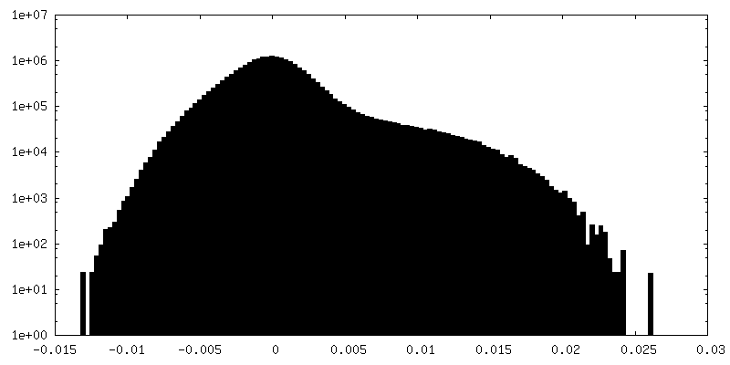
| 密度ヒストグラム |

- 試料の構成要素
-全体 : human heavy chain apoferritin
| 全体 | 名称: human heavy chain apoferritin |
|---|---|
| 要素 |
|
-超分子 #1: human heavy chain apoferritin
| 超分子 | 名称: human heavy chain apoferritin / タイプ: complex / キメラ: Yes / ID: 1 / 親要素: 0 / 含まれる分子: #1 |
|---|---|
| 由来(天然) | 生物種: Homo sapiens (ヒト) |
| 組換発現 | 生物種:  Escherichia coli BL21(DE3) (大腸菌) Escherichia coli BL21(DE3) (大腸菌) |
| 分子量 | 理論値: 20 KDa |
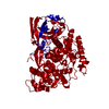
-分子 #1: Ferritin heavy chain
| 分子 | 名称: Ferritin heavy chain / タイプ: protein_or_peptide / ID: 1 / コピー数: 24 / 光学異性体: LEVO / EC番号: ferroxidase |
|---|---|
| 由来(天然) | 生物種: Homo sapiens (ヒト) |
| 分子量 | 理論値: 20.116547 KDa |
| 組換発現 | 生物種: Escherichia coli BL21(DE3) (大腸菌) |
| 配列 | 文字列: TSQVRQNYHQ DSEAAINRQI NLELYASYVY LSMSYYFDRD DVALKNFAKY FLHQSHEERE HAEKLMKLQN QRGGRIFLQD IKKPDCDDW ESGLNAMECA LHLEKNVNQS LLELHKLATD KNDPHLCDFI ETHYLNEQVK AIKELGDHVT NLRKMGAPES G LAEYLFDK HTLG |
-分子 #2: ZINC ION
| 分子 | 名称: ZINC ION / タイプ: ligand / ID: 2 / コピー数: 270 / 式: ZN |
|---|---|
| 分子量 | 理論値: 65.409 Da |
-分子 #3: SODIUM ION
| 分子 | 名称: SODIUM ION / タイプ: ligand / ID: 3 / コピー数: 112 |
|---|---|
| 分子量 | 理論値: 22.99 Da |
-分子 #4: water
| 分子 | 名称: water / タイプ: ligand / ID: 4 / コピー数: 2680 / 式: HOH |
|---|---|
| 分子量 | 理論値: 18.015 Da |
| Chemical component information |  ChemComp-HOH: |
-実験情報
-構造解析
| 手法 | クライオ電子顕微鏡法 |
|---|---|
 解析 解析 | 単粒子再構成法 |
| 試料の集合状態 | particle |
-試料調製
| 濃度 | 4.0 mg/mL | ||||||||||||
|---|---|---|---|---|---|---|---|---|---|---|---|---|---|
| 緩衝液 | pH: 7.5 構成要素:
| ||||||||||||
| グリッド | モデル: Quantifoil R2/2 / 材質: COPPER / メッシュ: 300 / 支持フィルム - 材質: CARBON / 支持フィルム - トポロジー: HOLEY / 支持フィルム - Film thickness: 5.0 nm / 前処理 - タイプ: GLOW DISCHARGE / 前処理 - 雰囲気: NITROGEN | ||||||||||||
| 凍結 | 凍結剤: ETHANE / チャンバー内湿度: 100 % / チャンバー内温度: 277.15 K / 装置: FEI VITROBOT MARK IV 詳細: 6 s blot time,30 s waiting time, ssDNA covered grid. |
- 電子顕微鏡法
| 顕微鏡 | FEI TITAN KRIOS |
|---|---|
| 電子線 | 加速電圧: 300 kV / 電子線源: FIELD EMISSION GUN |
| 電子光学系 | C2レンズ絞り径: 50.0 µm / 照射モード: FLOOD BEAM / 撮影モード: BRIGHT FIELDBright-field microscopy / Cs: 2.7 mm / 最大 デフォーカス(公称値): 1.7 µm / 最小 デフォーカス(公称値): 0.3 µm / 倍率(公称値): 130000 |
| 試料ステージ | 試料ホルダーモデル: FEI TITAN KRIOS AUTOGRID HOLDER ホルダー冷却材: NITROGEN |
| 撮影 | フィルム・検出器のモデル: FEI FALCON IV (4k x 4k) デジタル化 - サイズ - 横: 4096 pixel / デジタル化 - サイズ - 縦: 4096 pixel / デジタル化 - サンプリング間隔: 14.0 µm / 撮影したグリッド数: 1 / 実像数: 3282 / 平均露光時間: 4.08 sec. / 平均電子線量: 40.0 e/Å2 |
| 実験機器 |  モデル: Titan Krios / 画像提供: FEI Company |
-画像解析
| 粒子像選択 | 選択した数: 3282 |
|---|---|
| CTF補正 | ソフトウェア - 名称: CTFFIND (ver. 4) |
| 初期モデル | モデルのタイプ: EMDB MAP EMDB ID: |
| 初期 角度割当 | タイプ: MAXIMUM LIKELIHOOD / ソフトウェア - 名称: RELION (ver. 4.0) |
| 最終 3次元分類 | クラス数: 3 / ソフトウェア - 名称: RELION (ver. 4.0) |
| 最終 角度割当 | タイプ: MAXIMUM LIKELIHOOD / ソフトウェア - 名称: RELION (ver. 4.0) |
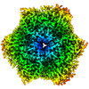
| 最終 再構成 | 使用したクラス数: 3 / 想定した対称性 - 点群: O (正8面体型対称) / アルゴリズム: BACK PROJECTION / 解像度のタイプ: BY AUTHOR / 解像度: 1.77 Å / 解像度の算出法: FSC 0.143 CUT-OFF / ソフトウェア - 名称: RELION (ver. 4.0) / 使用した粒子像数: 65786 |
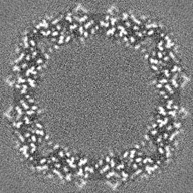
| FSC曲線 (解像度の算出) | 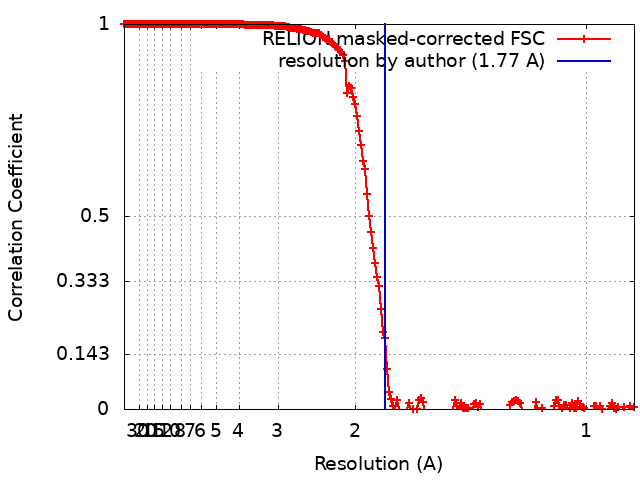 |


