 ムービー
ムービー コントローラー


+ データを開く

- 基本情報
基本情報
| 登録情報 | データベース: EMDB / ID: EMD-1467 | |||||||||
|---|---|---|---|---|---|---|---|---|---|---|
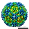
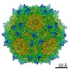
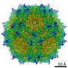
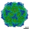
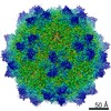
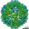
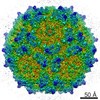
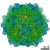
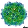
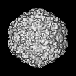
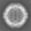
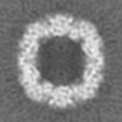
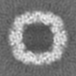
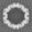
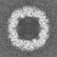
| タイトル | Human Parvovirus B19 (empty wildtype particle) | |||||||||
 マップデータ マップデータ | Human Parvovirus B19 (eB19, empty wildtype particle) | |||||||||
 試料 試料 |
| |||||||||
 キーワード キーワード |  icosahedral (二十面体) / empty viral particle / B19 icosahedral (二十面体) / empty viral particle / B19 | |||||||||
| 生物種 |  Human parvovirus B19 (B19 ウイルス) Human parvovirus B19 (B19 ウイルス) | |||||||||
| 手法 | 単粒子再構成法 / クライオ電子顕微鏡法 / 解像度: 11.3 Å | |||||||||
 データ登録者 データ登録者 | Kaufmann B / Chipman PR / Modrow S / Rossmann MG | |||||||||
 引用 引用 | ジャーナル: J Virol / 年: 2008 タイトル: Visualization of the externalized VP2 N termini of infectious human parvovirus B19. 著者: Bärbel Kaufmann / Paul R Chipman / Victor A Kostyuchenko / Susanne Modrow / Michael G Rossmann /  要旨: The structures of infectious human parvovirus B19 and empty wild-type particles were determined by cryoelectron microscopy (cryoEM) to 7.5-A and 11.3-A resolution, respectively, assuming icosahedral ...The structures of infectious human parvovirus B19 and empty wild-type particles were determined by cryoelectron microscopy (cryoEM) to 7.5-A and 11.3-A resolution, respectively, assuming icosahedral symmetry. Both of these, DNA filled and empty, wild-type particles contain a few copies of the minor capsid protein VP1. Comparison of wild-type B19 with the crystal structure and cryoEM reconstruction of recombinant B19 particles consisting of only the major capsid protein VP2 showed structural differences in the vicinity of the icosahedral fivefold axes. Although the unique N-terminal region of VP1 could not be visualized in the icosahedrally averaged maps, the N terminus of VP2 was shown to be exposed on the viral surface adjacent to the fivefold beta-cylinder. The conserved glycine-rich region is positioned between two neighboring, fivefold-symmetrically related VP subunits and not in the fivefold channel as observed for other parvoviruses. | |||||||||
| 履歴 |
|
- 構造の表示
構造の表示
| ムービー |
 ムービービューア ムービービューア |
|---|---|
| 構造ビューア | EMマップ: SurfViewMolmilJmol/JSmol |
| 添付画像 |

- ダウンロードとリンク
-EMDBアーカイブ
| マップデータ | emd_1467.map.gz | 2.5 MB | EMDBマップデータ形式 | |
|---|---|---|---|---|
| ヘッダ (付随情報) | emd-1467-v30.xmlemd-1467.xml | 10.3 KB 10.3 KB | 表示 表示 | EMDBヘッダ |
| 画像 |  1467.gif 1467.gif | 101.6 KB | ||
| アーカイブディレクトリ |  http://ftp.pdbj.org/pub/emdb/structures/EMD-1467ftp://ftp.pdbj.org/pub/emdb/structures/EMD-1467 http://ftp.pdbj.org/pub/emdb/structures/EMD-1467ftp://ftp.pdbj.org/pub/emdb/structures/EMD-1467 | HTTPS FTP |
-関連構造データ


-リンク
| EMDBのページ | EMDB (EBI/PDBe) / EMDataResource |
|---|
-マップ
| ファイル | ダウンロード / ファイル: emd_1467.map.gz / 形式: CCP4 / 大きさ: 5.1 MB / タイプ: IMAGE STORED AS FLOATING POINT NUMBER (4 BYTES) | ||||||||||||||||||||||||||||||||||||||||||||||||||||||||||||||||||||
|---|---|---|---|---|---|---|---|---|---|---|---|---|---|---|---|---|---|---|---|---|---|---|---|---|---|---|---|---|---|---|---|---|---|---|---|---|---|---|---|---|---|---|---|---|---|---|---|---|---|---|---|---|---|---|---|---|---|---|---|---|---|---|---|---|---|---|---|---|---|
| 注釈 | Human Parvovirus B19 (eB19, empty wildtype particle) | ||||||||||||||||||||||||||||||||||||||||||||||||||||||||||||||||||||
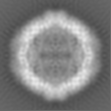
| 投影像・断面図 | 画像のコントロール
画像は Spider により作成 | ||||||||||||||||||||||||||||||||||||||||||||||||||||||||||||||||||||
| ボクセルのサイズ | X=Y=Z: 2.81649 Å | ||||||||||||||||||||||||||||||||||||||||||||||||||||||||||||||||||||
| 密度 |
| ||||||||||||||||||||||||||||||||||||||||||||||||||||||||||||||||||||
| 対称性 | 空間群: 1 | ||||||||||||||||||||||||||||||||||||||||||||||||||||||||||||||||||||
| 詳細 | EMDB XML:
CCP4マップ ヘッダ情報:
| ||||||||||||||||||||||||||||||||||||||||||||||||||||||||||||||||||||
 Z (Sec.)
 X (Row.)
 Y (Col.)


-添付データ
- 試料の構成要素
-全体 : Human Parvovirus B19 (empty wildtype particle)
| 全体 | 名称: Human Parvovirus B19 (empty wildtype particle) |
|---|---|
| 要素 |
|
-超分子 #1000: Human Parvovirus B19 (empty wildtype particle)
| 超分子 | 名称: Human Parvovirus B19 (empty wildtype particle) / タイプ: sample / ID: 1000 詳細: icosahedral, empty viral particle, non-enveloped protein shell 集合状態: 60 VP subunits form icosahedral protein shell / Number unique components: 1 |
|---|---|
| 分子量 | 理論値: 3.6 MDa |
-超分子 #1: Human parvovirus B19
| 超分子 | 名称: Human parvovirus B19 / タイプ: virus / ID: 1 / Name.synonym: Human Parvovirus B19 詳細: empty wildtype particles purified from human plasma, no ssDNA genome encapsidated NCBI-ID: 10798 / 生物種: Human parvovirus B19 / ウイルスタイプ: OTHER / ウイルス・単離状態: SPECIES / ウイルス・エンベロープ: No / ウイルス・中空状態: Yes / Syn species name: Human Parvovirus B19 |
|---|---|
| 宿主 | 生物種:  Homo sapiens (ヒト) / 別称: VERTEBRATES Homo sapiens (ヒト) / 別称: VERTEBRATES |
| 分子量 | 理論値: 3.6 MDa |
| ウイルス殻 | Shell ID: 1 / 直径: 260 Å / T番号(三角分割数): 1 |
-実験情報
-構造解析
| 手法 | クライオ電子顕微鏡法 |
|---|---|
 解析 解析 | 単粒子再構成法 |
| 試料の集合状態 | particle |
-試料調製
| 濃度 | 1 mg/mL |
|---|---|
| 緩衝液 | pH: 7.5 / 詳細: 25mM Tris-HCl |
| グリッド | 詳細: 400 mesh copper |
| 凍結 | 凍結剤: ETHANE / 装置: HOMEMADE PLUNGER 詳細: Vitrification instrument: guillotine-style plunge freezing device 手法: A small vial of ethane is placed inside a larger liquid nitrogen reservoir. The grid holding a few microliters of the sample is held in place at the bottom of a plunger by the means of fine ...手法: A small vial of ethane is placed inside a larger liquid nitrogen reservoir. The grid holding a few microliters of the sample is held in place at the bottom of a plunger by the means of fine tweezers. Once the ethane in the vial is completely frozen, it needs to be slightly melted. When the liquid ethane is ready, a piece of filter paper is then pressed against the sample to blot of excess buffer, sufficient to leave a thin layer on the grid. After a predetermined time, the filter paper is removed, and the plunger is allowed to drop into the liquid ethane. Once the grid enters the liquid ethane, the sample is rapidly frozen, and the grid is transferred under liquid nitrogen to a storage box immersed liquid nitrogen for later use in the microscope. |
- 電子顕微鏡法
| 顕微鏡 | FEI/PHILIPS CM300FEG/T |
|---|---|
| 電子線 | 加速電圧: 300 kV / 電子線源: FIELD EMISSION GUN |
| 電子光学系 | 倍率(補正後): 47000 / 照射モード: FLOOD BEAM / 撮影モード: BRIGHT FIELDBright-field microscopy / Cs: 2.0 mm / 最大 デフォーカス(公称値): 3.66 µm / 最小 デフォーカス(公称値): 1.25 µm / 倍率(公称値): 45000 |
| 試料ステージ | 試料ホルダー: Eucentric / 試料ホルダーモデル: GATAN LIQUID NITROGEN |
| 温度 | 平均: 98 K |
| アライメント法 | Legacy - 非点収差: live FFT at 200K |
| 詳細 | low dose |
| 撮影 | カテゴリ: FILM / フィルム・検出器のモデル: KODAK SO-163 FILM / デジタル化 - スキャナー: ZEISS SCAI / デジタル化 - サンプリング間隔: 14 µm / 実像数: 31 / 平均電子線量: 22 e/Å2 / ビット/ピクセル: 8 |
| Tilt angle min | 0 |
| Tilt angle max | 0 |
-画像解析
| CTF補正 | 詳細: each particle |
|---|---|
| 最終 再構成 | 想定した対称性 - 点群: I (正20面体型対称) / アルゴリズム: OTHER / 解像度のタイプ: BY AUTHOR / 解像度: 11.3 Å / 解像度の算出法: FSC 0.5 CUT-OFF / ソフトウェア - 名称: EMPFT, POR, P3DR 詳細: final map includes data to 10.5 Ang resolution (fsc 0.3 cut-off), magnification of final map standardized to a map calculated from B19 VP2 VLP model coordinates (PDB accession no 1S58) ...詳細: final map includes data to 10.5 Ang resolution (fsc 0.3 cut-off), magnification of final map standardized to a map calculated from B19 VP2 VLP model coordinates (PDB accession no 1S58) resulting in final pixel separation of 2.8165Ang 使用した粒子像数: 1959 |
| 詳細 | manual particle selection |
