 ムービー
ムービー コントローラー


+ データを開く

- 基本情報
基本情報
| 登録情報 | データベース: EMDB / ID: EMD-1334 | |||||||||
|---|---|---|---|---|---|---|---|---|---|---|
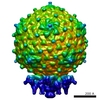
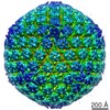
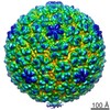
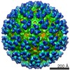
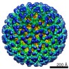
| タイトル | The structures of bacteriophages K1E and K1-5 explain processive degradation of polysaccharide capsules and evolution of new host specificities. | |||||||||
 マップデータ マップデータ | phage K1E icosahedrally averaged capsid | |||||||||
 試料 試料 |
| |||||||||
| 生物種 |  Enterobacteria phage K1E (ファージ) Enterobacteria phage K1E (ファージ) | |||||||||
| 手法 |  単粒子再構成法 / クライオ電子顕微鏡法 / ネガティブ染色法 / 解像度: 8.9 Å 単粒子再構成法 / クライオ電子顕微鏡法 / ネガティブ染色法 / 解像度: 8.9 Å | |||||||||
 データ登録者 データ登録者 | Leiman PG / Battisti AJ / Bowman VD / Stummeyer K / Muhlenhoff M / Gerardy-Schahn R / Scholl D / Molineux IJ | |||||||||
 引用 引用 | ジャーナル: J Mol Biol / 年: 2007 タイトル: The structures of bacteriophages K1E and K1-5 explain processive degradation of polysaccharide capsules and evolution of new host specificities. 著者: Petr G Leiman / Anthony J Battisti / Valorie D Bowman / Katharina Stummeyer / Martina Mühlenhoff / Rita Gerardy-Schahn / Dean Scholl / Ian J Molineux /  要旨: External polysaccharides of many pathogenic bacteria form capsules protecting the bacteria from the animal immune system and phage infection. However, some bacteriophages can digest these capsules ...External polysaccharides of many pathogenic bacteria form capsules protecting the bacteria from the animal immune system and phage infection. However, some bacteriophages can digest these capsules using glycosidases displayed on the phage particle. We have utilized cryo-electron microscopy to determine the structures of phages K1E and K1-5 and thereby establish the mechanism by which these phages attain and switch their host specificity. Using a specific glycosidase, both phages penetrate the capsule and infect the neuroinvasive human pathogen Escherichia coli K1. In addition to the K1-specific glycosidase, each K1-5 particle carries a second enzyme that allows it to infect E. coli K5, whose capsule is chemically different from that of K1. The enzymes are organized into a multiprotein complex attached via an adapter protein to the virus portal vertex, through which the DNA is ejected during infection. The structure of the complex suggests a mechanism for the apparent processivity of degradation that occurs as the phage drills through the polysaccharide capsule. The enzymes recognize the adapter protein by a conserved N-terminal sequence, providing a mechanism for phages to acquire different enzymes and thus to evolve new host specificities. | |||||||||
| 履歴 |
|
- 構造の表示
構造の表示
| ムービー |
 ムービービューア ムービービューア |
|---|---|
| 構造ビューア | EMマップ: SurfViewMolmilJmol/JSmol |
| 添付画像 |

- ダウンロードとリンク
-EMDBアーカイブ
| マップデータ | emd_1334.map.gz | 32.2 MB | EMDBマップデータ形式 | |
|---|---|---|---|---|
| ヘッダ (付随情報) | emd-1334-v30.xmlemd-1334.xml | 10 KB 10 KB | 表示 表示 | EMDBヘッダ |
| 画像 | 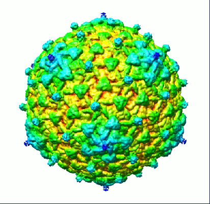 1334.gif 1334.gif | 79.9 KB | ||
| アーカイブディレクトリ |  http://ftp.pdbj.org/pub/emdb/structures/EMD-1334ftp://ftp.pdbj.org/pub/emdb/structures/EMD-1334 http://ftp.pdbj.org/pub/emdb/structures/EMD-1334ftp://ftp.pdbj.org/pub/emdb/structures/EMD-1334 | HTTPS FTP |
-関連構造データ
-リンク
| EMDBのページ | EMDB (EBI/PDBe) / EMDataResource |
|---|
-マップ
| ファイル | ダウンロード / ファイル: emd_1334.map.gz / 形式: CCP4 / 大きさ: 96.6 MB / タイプ: IMAGE STORED AS FLOATING POINT NUMBER (4 BYTES) | ||||||||||||||||||||||||||||||||||||||||||||||||||||||||||||||||||||
|---|---|---|---|---|---|---|---|---|---|---|---|---|---|---|---|---|---|---|---|---|---|---|---|---|---|---|---|---|---|---|---|---|---|---|---|---|---|---|---|---|---|---|---|---|---|---|---|---|---|---|---|---|---|---|---|---|---|---|---|---|---|---|---|---|---|---|---|---|---|
| 注釈 | phage K1E icosahedrally averaged capsid | ||||||||||||||||||||||||||||||||||||||||||||||||||||||||||||||||||||
| ボクセルのサイズ | X=Y=Z: 2.98 Å | ||||||||||||||||||||||||||||||||||||||||||||||||||||||||||||||||||||
| 密度 |
| ||||||||||||||||||||||||||||||||||||||||||||||||||||||||||||||||||||
| 対称性 | 空間群: 1 | ||||||||||||||||||||||||||||||||||||||||||||||||||||||||||||||||||||
| 詳細 | EMDB XML:
CCP4マップ ヘッダ情報:
| ||||||||||||||||||||||||||||||||||||||||||||||||||||||||||||||||||||
-添付データ
- 試料の構成要素
-全体 : Bacteriophages K1E and K1-5
| 全体 | 名称: Bacteriophages K1E and K1-5 |
|---|---|
| 要素 |
|
-超分子 #1000: Bacteriophages K1E and K1-5
| 超分子 | 名称: Bacteriophages K1E and K1-5 / タイプ: sample / ID: 1000 詳細: The tail and internal capsid proteins are not resolved in the map because the particle was icosahedrally averaged. 集合状態: complete virus particle / Number unique components: 1 |
|---|---|
| 分子量 | 実験値: 48 MDa / 理論値: 58 MDa / 手法: Primary sequence |
-超分子 #1: Enterobacteria phage K1E
| 超分子 | 名称: Enterobacteria phage K1E / タイプ: virus / ID: 1 / Name.synonym: phage K1E / 詳細: complete virus particle / NCBI-ID: 344022 / 生物種: Enterobacteria phage K1E / ウイルスタイプ: VIRION / ウイルス・単離状態: SPECIES / ウイルス・エンベロープ: No / ウイルス・中空状態: No / Syn species name: phage K1E |
|---|---|
| 宿主 | 生物種:  Escherichia coli (大腸菌) / 別称: BACTERIA(EUBACTERIA) Escherichia coli (大腸菌) / 別称: BACTERIA(EUBACTERIA) |
| 分子量 | 実験値: 58 MDa / 理論値: 58 MDa |
| ウイルス殻 | Shell ID: 1 / 名称: coat protein / 直径: 310 Å / T番号(三角分割数): 7 |
-実験情報
-構造解析
| 手法 | ネガティブ染色法, クライオ電子顕微鏡法 |
|---|---|
 解析 解析 | 単粒子再構成法 |
| 試料の集合状態 | particle |
-試料調製
| 濃度 | 1 mg/mL |
|---|---|
| 緩衝液 | pH: 7.5 / 詳細: 50 mM Tris HCl pH 7.5, 100 mM NaCl, 8 mM MgSO4 |
| 染色 | タイプ: NEGATIVE / 詳細: no staining |
| グリッド | 詳細: quantifoil grid |
| 凍結 | 凍結剤: ETHANE / チャンバー内湿度: 80 % / チャンバー内温度: 300 K / 装置: OTHER / 詳細: Vitrification instrument: Vitrobot |
- 電子顕微鏡法
| 顕微鏡 | FEI/PHILIPS CM300FEG/T |
|---|---|
| 電子線 | 加速電圧: 300 kV / 電子線源: FIELD EMISSION GUN |
| 電子光学系 | 倍率(補正後): 47000 / 照射モード: SPOT SCAN / 撮影モード: BRIGHT FIELDBright-field microscopy / Cs: 2.0 mm / 最大 デフォーカス(公称値): 3.3 µm / 最小 デフォーカス(公称値): 0.7 µm / 倍率(公称値): 45000 |
| 試料ステージ | 試料ホルダー: Side entry liquid nitrogen-cooled cryo specimen holder 試料ホルダーモデル: GATAN LIQUID NITROGEN |
| 温度 | 平均: 100 K |
| 日付 | 2005年5月10日 |
| 撮影 | カテゴリ: FILM / フィルム・検出器のモデル: KODAK SO-163 FILM / デジタル化 - スキャナー: ZEISS SCAI / デジタル化 - サンプリング間隔: 7 µm / 実像数: 122 / 平均電子線量: 25 e/Å2 / ビット/ピクセル: 12 |
| Tilt angle min | 0 |
| Tilt angle max | 0 |
-画像解析
| CTF補正 | 詳細: Each particle |
|---|---|
| 最終 2次元分類 | クラス数: 296 |
| 最終 再構成 | 想定した対称性 - 点群: I (正20面体型対称) / アルゴリズム: OTHER / 解像度のタイプ: BY AUTHOR / 解像度: 8.9 Å / 解像度の算出法: OTHER / ソフトウェア - 名称: EMAN / 使用した粒子像数: 6105 |
