 ムービー
ムービー コントローラー


+ データを開く

- 基本情報
基本情報
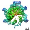
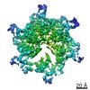
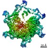
| 登録情報 | データベース: EMDB / ID: EMD-1218 | |||||||||
|---|---|---|---|---|---|---|---|---|---|---|
| タイトル | The structural basis for regulated assembly and function of the transcriptional activator NtrC. | |||||||||
 マップデータ マップデータ | 3D map of full-length, BeF-activated NtrC in the ADP-AlF state | |||||||||
 試料 試料 |
| |||||||||
| 生物種 |   Salmonella enterica subsp. enterica serovar Typhimurium (サルモネラ菌) Salmonella enterica subsp. enterica serovar Typhimurium (サルモネラ菌) | |||||||||
| 手法 | 単粒子再構成法 / ネガティブ染色法 / 解像度: 28.0 Å | |||||||||
 データ登録者 データ登録者 | De Carlo S / Chen B / Hoover TR / Kondrashkina E / Nogales E / Nixon BT | |||||||||
 引用 引用 | ジャーナル: Genes Dev / 年: 2006 タイトル: The structural basis for regulated assembly and function of the transcriptional activator NtrC. 著者: Sacha De Carlo / Baoyu Chen / Timothy R Hoover / Elena Kondrashkina / Eva Nogales / B Tracy Nixon /  要旨: In two-component signal transduction, an input triggers phosphorylation of receiver domains that regulate the status of output modules. One such module is the AAA+ ATPase domain in bacterial enhancer- ...In two-component signal transduction, an input triggers phosphorylation of receiver domains that regulate the status of output modules. One such module is the AAA+ ATPase domain in bacterial enhancer-binding proteins that remodel the sigma(54) form of RNA polymerase. We report X-ray solution scattering and electron microscopy structures of the activated, full-length nitrogen-regulatory protein C (NtrC) showing a novel mechanism for regulation of AAA+ ATPase assembly via the juxtaposition of the receiver domains and ATPase ring. Accompanying the hydrolysis cycle that is required for transcriptional activation, we observed major order-disorder changes in the GAFTGA loops involved in sigma(54) binding, as well as in the DNA-binding domains. | |||||||||
| 履歴 |
|
- 構造の表示
構造の表示
| ムービー |
 ムービービューア ムービービューア |
|---|---|
| 構造ビューア | EMマップ: SurfViewMolmilJmol/JSmol |
| 添付画像 |

- ダウンロードとリンク
-EMDBアーカイブ
| マップデータ | emd_1218.map.gz | 5.2 MB | EMDBマップデータ形式 | |
|---|---|---|---|---|
| ヘッダ (付随情報) | emd-1218-v30.xmlemd-1218.xml | 9.1 KB 9.1 KB | 表示 表示 | EMDBヘッダ |
| 画像 | 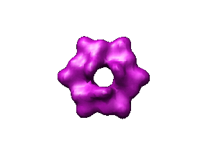 1218.gif 1218.gif | 9.5 KB | ||
| アーカイブディレクトリ |  http://ftp.pdbj.org/pub/emdb/structures/EMD-1218ftp://ftp.pdbj.org/pub/emdb/structures/EMD-1218 http://ftp.pdbj.org/pub/emdb/structures/EMD-1218ftp://ftp.pdbj.org/pub/emdb/structures/EMD-1218 | HTTPS FTP |
-関連構造データ
-リンク
| EMDBのページ | EMDB (EBI/PDBe) / EMDataResource |
|---|
-マップ
| ファイル | ダウンロード / ファイル: emd_1218.map.gz / 形式: CCP4 / 大きさ: 7.3 MB / タイプ: IMAGE STORED AS FLOATING POINT NUMBER (4 BYTES) | ||||||||||||||||||||||||||||||||||||||||||||||||||||||||||||||||||||
|---|---|---|---|---|---|---|---|---|---|---|---|---|---|---|---|---|---|---|---|---|---|---|---|---|---|---|---|---|---|---|---|---|---|---|---|---|---|---|---|---|---|---|---|---|---|---|---|---|---|---|---|---|---|---|---|---|---|---|---|---|---|---|---|---|---|---|---|---|---|
| 注釈 | 3D map of full-length, BeF-activated NtrC in the ADP-AlF state | ||||||||||||||||||||||||||||||||||||||||||||||||||||||||||||||||||||
| ボクセルのサイズ | X=Y=Z: 2.56 Å | ||||||||||||||||||||||||||||||||||||||||||||||||||||||||||||||||||||
| 密度 |
| ||||||||||||||||||||||||||||||||||||||||||||||||||||||||||||||||||||
| 対称性 | 空間群: 1 | ||||||||||||||||||||||||||||||||||||||||||||||||||||||||||||||||||||
| 詳細 | EMDB XML:
CCP4マップ ヘッダ情報:
| ||||||||||||||||||||||||||||||||||||||||||||||||||||||||||||||||||||
-添付データ
- 試料の構成要素
-全体 : Full length, active NtrC
| 全体 | 名称: Full length, active NtrCFull Length LP |
|---|---|
| 要素 |
|
-超分子 #1000: Full length, active NtrC
| 超分子 | 名称: Full length, active NtrC / タイプ: sample / ID: 1000 / 詳細: Activated with Mg/BeF / 集合状態: Hexamer / Number unique components: 1 |
|---|---|
| 分子量 | 実験値: 300 KDa / 理論値: 300 KDa / 手法: Sedimentation Velocity, Analytical centrifugation |
-分子 #1: Transcription activator
| 分子 | 名称: Transcription activator / タイプ: protein_or_peptide / ID: 1 / Name.synonym: Enhancer-binding protein 詳細: Baers following mutations: S160F, R456A, N457A, R461A コピー数: 1 / 集合状態: Hexamer / 組換発現: Yes |
|---|---|
| 由来(天然) | 生物種: Salmonella enterica subsp. enterica serovar Typhimurium (サルモネラ菌) 別称: Bacteria / 細胞: Salmonella typhimurium |
| 分子量 | 実験値: 300 KDa / 理論値: 300 KDa |
| 組換発現 | 生物種: Escherichia coli (大腸菌) |
-実験情報
-構造解析
| 手法 | ネガティブ染色法 |
|---|---|
 解析 解析 | 単粒子再構成法 |
| 試料の集合状態 | particle |
-試料調製
| 濃度 | 0.05 mg/mL |
|---|---|
| 緩衝液 | pH: 8.2 詳細: 200mM KCl, 20mM Tris, 1mM nucleotide, 0.2 mM BeCl2 and 5mM NaF, 5mM MgCl2, 5% Trehalose |
| 染色 | タイプ: NEGATIVE / 詳細: 3% uranyl acetate |
| 凍結 | 凍結剤: NONE |
- 電子顕微鏡法
| 顕微鏡 | FEI TECNAI 12 |
|---|---|
| 電子線 | 加速電圧: 120 kV / 電子線源: LAB6 |
| 電子光学系 | 倍率(補正後): 49767 / 照射モード: FLOOD BEAM / 撮影モード: BRIGHT FIELDBright-field microscopy / Cs: 6.2 mm / 最大 デフォーカス(公称値): 1.5 µm / 最小 デフォーカス(公称値): 0.9 µm / 倍率(公称値): 50000 |
| 試料ステージ | 試料ホルダー: simple tilt / 試料ホルダーモデル: OTHER / Tilt angle max: 50 |
| アライメント法 | Legacy - 非点収差: corrected / Legacy - Electron beam tilt params: 0 |
| 撮影 | カテゴリ: FILM / フィルム・検出器のモデル: KODAK SO-163 FILM / デジタル化 - スキャナー: OTHER / デジタル化 - サンプリング間隔: 6.7 µm / 実像数: 18 / 平均電子線量: 20 e/Å2 / ビット/ピクセル: 14 |
| Tilt angle min | 0 |
-画像解析
| 最終 再構成 | 想定した対称性 - 点群: C3 (3回回転対称) / アルゴリズム: OTHER / 解像度のタイプ: BY AUTHOR / 解像度: 28.0 Å / 解像度の算出法: FSC 0.5 CUT-OFF / ソフトウェア - 名称: SPIDER / 使用した粒子像数: 3500 |
|---|
