Type-1 KH domain profile. / Ribosomal protein S21, conserved site / Ribosomal protein S21 signature. / Ribosomal protein S14, bacterial/plastid / Ribosomal protein S21 superfamily / Ribosomal protein S21 / Ribosomal protein S21 / Ribosomal protein S3, bacterial-type / Ribosomal protein S6, conserved site / Ribosomal protein S6 signature. ...Type-1 KH domain profile. / Ribosomal protein S21, conserved site / Ribosomal protein S21 signature. / Ribosomal protein S14, bacterial/plastid / Ribosomal protein S21 superfamily / Ribosomal protein S21 / Ribosomal protein S21 / Ribosomal protein S3, bacterial-type / Ribosomal protein S6, conserved site / Ribosomal protein S6 signature. / Ribosomal protein S19, bacterial-type / Ribosomal protein S7, bacterial/organellar-type / Ribosomal protein S11, bacterial-type / Ribosomal protein S13, bacterial-type / Ribosomal protein S20 / Ribosomal protein S20 superfamily / Ribosomal protein S20 / Ribosomal protein S9, bacterial/plastid / Ribosomal protein S4, bacterial-type / 30S ribosomal protein S17 / Ribosomal protein S5, bacterial-type / Ribosomal protein S6, plastid/chloroplast / Ribosomal protein S2, bacteria/mitochondria/plastid / Ribosomal protein S18, conserved site / Ribosomal protein S18 signature. / Ribosomal protein S16 / Ribosomal protein S16 / Ribosomal protein S16 domain superfamily / Ribosomal protein S15, bacterial-type / Ribosomal protein S2 signature 2. / Ribosomal protein S6 / Ribosomal protein S6 / Ribosomal protein S6 superfamily / Ribosomal protein S12, bacterial-type / Translation elongation factor EF1B/ribosomal protein S6 / Ribosomal protein S18 / Ribosomal protein S18 / Ribosomal protein S18 superfamily / KHドメイン / K homology RNA-binding domain / Ribosomal protein S3, conserved site / Ribosomal protein S14, conserved site / Ribosomal protein S10, conserved site / : / K Homology domain, type 2 / Ribosomal protein S3, C-terminal / Ribosomal protein S3, C-terminal domain superfamily / Ribosomal protein S15/S19, conserved site / KHドメイン / Ribosomal protein S19/S15 / Ribosomal protein S19/S15, superfamily / Ribosomal protein S10 / Ribosomal protein S3, C-terminal domain / Ribosomal protein S3 signature. / Ribosomal protein S10 signature. / Ribosomal protein S14 signature. / Ribosomal protein S7, conserved site / K homology domain superfamily, prokaryotic type / Ribosomal protein S19 / Ribosomal protein S2 signature 1. / Ribosomal protein S13, conserved site / Ribosomal protein S2, conserved site / Ribosomal protein S13 / 30s ribosomal protein S13, C-terminal / Ribosomal protein S2 / Ribosomal protein S2, flavodoxin-like domain superfamily / Ribosomal protein S14 / Ribosomal protein S2 / Ribosomal protein S4/S9 N-terminal domain / Type-2 KH domain profile. / Ribosomal protein S4/S9, N-terminal / Ribosomal protein S4, conserved site / Ribosomal protein S4/S9 N-terminal domain / Ribosomal protein S13/S18 / Ribosomal protein S4/S9 / Ribosomal protein S19 signature. / K homology domain-like, alpha/beta / Ribosomal protein S14p/S29e / Ribosomal protein S8 / Ribosomal protein S8 superfamily / Ribosomal protein S5, N-terminal, conserved site / Ribosomal protein S5 signature. / Ribosomal protein S7 signature. / Ribosomal protein S5 / Ribosomal protein S5, N-terminal / Ribosomal S11, conserved site / Ribosomal protein S10p/S20e / Ribosomal protein S13-like, H2TH / S5 double stranded RNA-binding domain profile. / Ribosomal protein S5, C-terminal / Ribosomal protein S9, conserved site / Ribosomal protein S5, N-terminal domain / Ribosomal protein S8 / Ribosomal protein S10 domain / Ribosomal protein S10 domain superfamily / Ribosomal protein S5, C-terminal domain / S4 RNA-binding domain / Ribosomal protein S11 / RNA-binding S4 domain / Ribosomal protein S13 signature. 類似検索 - ドメイン・相同性
Small ribosomal subunit protein uS9 / Small ribosomal subunit protein bS6 / Small ribosomal subunit protein bS21 / Small ribosomal subunit protein uS12 / Small ribosomal subunit protein uS15 / Small ribosomal subunit protein uS4 / Small ribosomal subunit protein bS20 / Small ribosomal subunit protein uS2 / Small ribosomal subunit protein uS3 / Small ribosomal subunit protein uS5 ...Small ribosomal subunit protein uS9 / Small ribosomal subunit protein bS6 / Small ribosomal subunit protein bS21 / Small ribosomal subunit protein uS12 / Small ribosomal subunit protein uS15 / Small ribosomal subunit protein uS4 / Small ribosomal subunit protein bS20 / Small ribosomal subunit protein uS2 / Small ribosomal subunit protein uS3 / Small ribosomal subunit protein uS5 / Small ribosomal subunit protein bS18 / Small ribosomal subunit protein uS7 / Small ribosomal subunit protein bS16 / Small ribosomal subunit protein uS10 / Small ribosomal subunit protein uS19 / Small ribosomal subunit protein uS17 / Small ribosomal subunit protein uS14 / Small ribosomal subunit protein uS8 / Small ribosomal subunit protein uS13 / Small ribosomal subunit protein uS11 類似検索 - 構成要素
iNEXT, project number 653706, funded by the Horizon 2020 programme of the European Union
引用
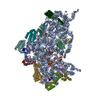
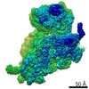
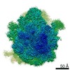
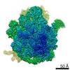
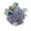
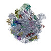
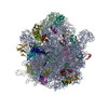
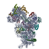
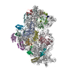
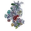
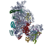
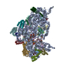
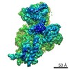
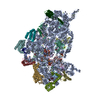
ジャーナル: Proc Natl Acad Sci U S A / 年: 2019 タイトル: Structure of ribosomes from an aminoglycoside-resistant clinical isolate. 著者: Yehuda Halfon / Alicia Jimenez-Fernandez / Ruggero La Rosa / Rocio Espinosa Portero / Helle Krogh Johansen / Donna Matzov / Zohar Eyal / Anat Bashan / Ella Zimmerman / Matthew Belousoff / ...著者: Yehuda Halfon / Alicia Jimenez-Fernandez / Ruggero La Rosa / Rocio Espinosa Portero / Helle Krogh Johansen / Donna Matzov / Zohar Eyal / Anat Bashan / Ella Zimmerman / Matthew Belousoff / Søren Molin / Ada Yonath / 要旨: Resistance to antibiotics has become a major threat to modern medicine. The ribosome plays a fundamental role in cell vitality by the translation of the genetic code into proteins; hence, it is a ...Resistance to antibiotics has become a major threat to modern medicine. The ribosome plays a fundamental role in cell vitality by the translation of the genetic code into proteins; hence, it is a major target for clinically useful antibiotics. We report here the cryo-electron microscopy structures of the ribosome of a pathogenic aminoglycoside (AG)-resistant strain, as well as of a nonresistance strain isolated from a cystic fibrosis patient. The structural studies disclosed defective ribosome complex formation due to a conformational change of rRNA helix H69, an essential intersubunit bridge, and a secondary binding site of the AGs. In addition, a stable conformation of nucleotides A1486 and A1487, pointing into helix h44, is created compared to a non-AG-bound ribosome. We suggest that altering the conformations of ribosomal protein uL6 and rRNA helix H69, which interact with initiation-factor IF2, interferes with proper protein synthesis initiation.
 ムービー
ムービー コントローラー


 データを開く
 基本情報
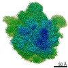
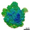
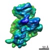
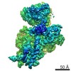
基本情報 マップデータ
 試料
試料 機能・相同性情報
 tRNA binding /

 データ登録者
 デンマーク, 4件
  引用
引用

 構造の表示
構造の表示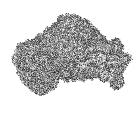
 ダウンロードとリンク
 emd_10281.png
 http://ftp.pdbj.org/pub/emdb/structures/EMD-10281


 試料の構成要素
 解析
解析 電子顕微鏡法