 ムービー
ムービー コントローラー


+ データを開く

- 基本情報
基本情報
| 登録情報 | データベース: EMDB / ID: EMD-6335 | |||||||||
|---|---|---|---|---|---|---|---|---|---|---|
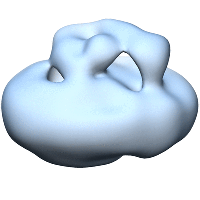
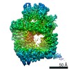
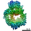
| タイトル | Three-Dimensional Reconstruction of Lipid Nanodisc Reconstituted Yeast V-ATPase Membrane Sector | |||||||||
 マップデータ マップデータ | Reconstruction of Lipid Nanodisc Reconstituted Yeast V-ATPase Membrane Sector | |||||||||
 試料 試料 |
| |||||||||
| 生物種 |   Saccharomyces cerevisiae (パン酵母) / synthetic construct (人工物) Saccharomyces cerevisiae (パン酵母) / synthetic construct (人工物) | |||||||||
| 手法 | 単粒子再構成法 / ネガティブ染色法 / 解像度: 16.3 Å | |||||||||
 データ登録者 データ登録者 | Stam NJ / Wilkens S | |||||||||
 引用 引用 | ジャーナル: J Biol Chem / 年: 2017 タイトル: Structure of the Lipid Nanodisc-reconstituted Vacuolar ATPase Proton Channel: DEFINITION OF THE INTERACTION OF ROTOR AND STATOR AND IMPLICATIONS FOR ENZYME REGULATION BY REVERSIBLE DISSOCIATION. 著者: Nicholas J Stam / Stephan Wilkens /  要旨: Eukaryotic vacuolar H-ATPase (V-ATPase) is a multisubunit enzyme complex that acidifies subcellular organelles and the extracellular space. V-ATPase consists of soluble V-ATPase and membrane-integral ...Eukaryotic vacuolar H-ATPase (V-ATPase) is a multisubunit enzyme complex that acidifies subcellular organelles and the extracellular space. V-ATPase consists of soluble V-ATPase and membrane-integral V proton channel sectors. To investigate the mechanism of V-ATPase regulation by reversible disassembly, we recently determined a cryo-EM reconstruction of yeast V The structure indicated that, when V is released from V, the N-terminal cytoplasmic domain of subunit a (a) changes conformation to bind rotor subunit d However, insufficient resolution precluded a precise definition of the a-d interface. Here we reconstituted V into lipid nanodiscs for single-particle EM. 3D reconstructions calculated at ∼15-Å resolution revealed two sites of contact between a and d that are mediated by highly conserved charged residues. Alanine mutagenesis of some of these residues disrupted the a-d interaction, as shown by isothermal titration calorimetry and gel filtration of recombinant subunits. A recent cryo-EM study of holo V-ATPase revealed three major conformations corresponding to three rotational states of the central rotor of the enzyme. Comparison of the three V-ATPase conformations with the structure of nanodisc-bound V revealed that V is halted in rotational state 3. Combined with our prior work that showed autoinhibited V-ATPase to be arrested in state 2, we propose a model in which the conformational mismatch between free V and V functions to prevent unintended reassembly of holo V-ATPase when activity is not needed. | |||||||||
| 履歴 |
|
- 構造の表示
構造の表示
| ムービー |
 ムービービューア ムービービューア |
|---|---|
| 構造ビューア | EMマップ: SurfViewMolmilJmol/JSmol |
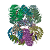
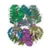
| 添付画像 |
- ダウンロードとリンク
-EMDBアーカイブ
| マップデータ | emd_6335.map.gz | 312.3 KB | EMDBマップデータ形式 | |
|---|---|---|---|---|
| ヘッダ (付随情報) | emd-6335-v30.xmlemd-6335.xml | 9.6 KB 9.6 KB | 表示 表示 | EMDBヘッダ |
| FSC (解像度算出) | emd_6335_fsc.xml | 4.1 KB | 表示 | FSCデータファイル |
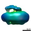
| 画像 |  400_6335.gif 400_6335.gif 80_6335.gif 80_6335.gif | 57.7 KB 12.4 KB | ||
| アーカイブディレクトリ |  http://ftp.pdbj.org/pub/emdb/structures/EMD-6335ftp://ftp.pdbj.org/pub/emdb/structures/EMD-6335 http://ftp.pdbj.org/pub/emdb/structures/EMD-6335ftp://ftp.pdbj.org/pub/emdb/structures/EMD-6335 | HTTPS FTP |
-関連構造データ

-リンク
| EMDBのページ | EMDB (EBI/PDBe) / EMDataResource |
|---|
-マップ
| ファイル | ダウンロード / ファイル: emd_6335.map.gz / 形式: CCP4 / 大きさ: 3.3 MB / タイプ: IMAGE STORED AS FLOATING POINT NUMBER (4 BYTES) | ||||||||||||||||||||||||||||||||||||||||||||||||||||||||||||
|---|---|---|---|---|---|---|---|---|---|---|---|---|---|---|---|---|---|---|---|---|---|---|---|---|---|---|---|---|---|---|---|---|---|---|---|---|---|---|---|---|---|---|---|---|---|---|---|---|---|---|---|---|---|---|---|---|---|---|---|---|---|
| 注釈 | Reconstruction of Lipid Nanodisc Reconstituted Yeast V-ATPase Membrane Sector | ||||||||||||||||||||||||||||||||||||||||||||||||||||||||||||
| ボクセルのサイズ | X=Y=Z: 3.5 Å | ||||||||||||||||||||||||||||||||||||||||||||||||||||||||||||
| 密度 |
| ||||||||||||||||||||||||||||||||||||||||||||||||||||||||||||
| 対称性 | 空間群: 1 | ||||||||||||||||||||||||||||||||||||||||||||||||||||||||||||
| 詳細 | EMDB XML:
CCP4マップ ヘッダ情報:
| ||||||||||||||||||||||||||||||||||||||||||||||||||||||||||||
-添付データ
- 試料の構成要素
-全体 : V-ATPase membrane sector (Vo) isolated from yeast membranes and r...
| 全体 | 名称: V-ATPase membrane sector (Vo) isolated from yeast membranes and reconstituted with lipid nanodisc |
|---|---|
| 要素 |
|
-超分子 #1000: V-ATPase membrane sector (Vo) isolated from yeast membranes and r...
| 超分子 | 名称: V-ATPase membrane sector (Vo) isolated from yeast membranes and reconstituted with lipid nanodisc タイプ: sample / ID: 1000 / Number unique components: 2 |
|---|---|
| 分子量 | 理論値: 400 KDa |
-分子 #1: Vacuolar-type (V-) ATPase membrane sector (Vo)
| 分子 | 名称: Vacuolar-type (V-) ATPase membrane sector (Vo) / タイプ: protein_or_peptide / ID: 1 / 組換発現: No / データベース: NCBI |
|---|---|
| 由来(天然) | 生物種: Saccharomyces cerevisiae (パン酵母) / 株: BY4743 / 別称: Yeast |
| 分子量 | 理論値: 300 KDa |
-分子 #2: MSP1E3D1
| 分子 | 名称: MSP1E3D1 / タイプ: protein_or_peptide / ID: 2 / Name.synonym: membrane scaffold protein, nanodisc / コピー数: 2 / 集合状態: Dimer / 組換発現: Yes |
|---|---|
| 由来(天然) | 生物種: synthetic construct (人工物) |
| 分子量 | 理論値: 30 KDa |
| 組換発現 | 生物種:  Escherichia coli BL21(DE3) (大腸菌) / 組換株: BL21(DE3) / 組換プラスミド: pET28a Escherichia coli BL21(DE3) (大腸菌) / 組換株: BL21(DE3) / 組換プラスミド: pET28a |
-実験情報
-構造解析
| 手法 | ネガティブ染色法 |
|---|---|
 解析 解析 | 単粒子再構成法 |
| 試料の集合状態 | particle |
-試料調製
| 濃度 | 0.04 mg/mL |
|---|---|
| 緩衝液 | pH: 7.4 / 詳細: 20 mM Tris-HCl, 100 mM NaCl, 0.5 mM EDTA |
| 染色 | タイプ: NEGATIVE 詳細: 2% w/v uranyl formate applied to grids with adsorbed protein |
| グリッド | 詳細: 200 mesh copper grid with thin carbon support, glow-discharged in air |
| 凍結 | 凍結剤: NONE / 装置: OTHER |
- 電子顕微鏡法
| 顕微鏡 | JEOL 2100 |
|---|---|
| 電子線 | 加速電圧: 200 kV / 電子線源: LAB6 |
| 電子光学系 | 倍率(補正後): 85800 / 照射モード: FLOOD BEAM / 撮影モード: BRIGHT FIELDBright-field microscopy / Cs: 2.0 mm / 最大 デフォーカス(公称値): 2.08 µm / 最小 デフォーカス(公称値): 1.125 µm / 倍率(公称値): 60000 |
| 試料ステージ | 試料ホルダーモデル: JEOL |
| アライメント法 | Legacy - 非点収差: Objective lens astigmatism was corrected at 100,000 times magnification. |
| 日付 | 2014年11月13日 |
| 撮影 | カテゴリ: CCD フィルム・検出器のモデル: TVIPS TEMCAM-F415 (4k x 4k) 実像数: 390 / 平均電子線量: 17 e/Å2 / ビット/ピクセル: 8 |
-画像解析
| CTF補正 | 詳細: By micrograph |
|---|---|
| 最終 再構成 | アルゴリズム: OTHER / 解像度のタイプ: BY AUTHOR / 解像度: 16.3 Å / 解像度の算出法: OTHER / ソフトウェア - 名称: EMAN2, Relion / 使用した粒子像数: 47422 |
| 詳細 | Particles were selected using a monitored semi-automated selection program. |
| FSC曲線 (解像度の算出) | 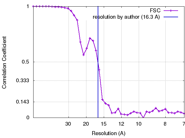 |
