ムービー
ムービー コントローラー


+ データを開く

- 基本情報
基本情報
| 登録情報 | データベース: PDB / ID: 5ide | ||||||
|---|---|---|---|---|---|---|---|
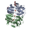
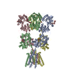
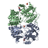
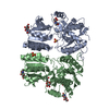
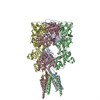
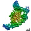
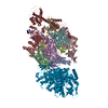
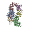
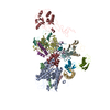
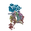
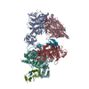
| タイトル | Cryo-EM structure of GluA2/3 AMPA receptor heterotetramer (model I) | ||||||
 要素 要素 |
| ||||||
 キーワード キーワード |  SIGNALING PROTEIN / AMPA glutamate receptor SIGNALING PROTEIN / AMPA glutamate receptor | ||||||
| 機能・相同性 |  機能・相同性情報 機能・相同性情報chemical synaptic transmission, postsynaptic / Trafficking of AMPA receptors / Synaptic adhesion-like molecules / parallel fiber to Purkinje cell synapse / spine synapse / dendritic spine neck / dendritic spine head / Activation of AMPA receptors / response to lithium ion / perisynaptic space ...chemical synaptic transmission, postsynaptic / Trafficking of AMPA receptors / Synaptic adhesion-like molecules / parallel fiber to Purkinje cell synapse / spine synapse / dendritic spine neck / dendritic spine head / Activation of AMPA receptors / response to lithium ion / perisynaptic space / cellular response to glycine / AMPA glutamate receptor activity / Trafficking of GluR2-containing AMPA receptors / immunoglobulin binding / AMPA glutamate receptor complex / kainate selective glutamate receptor activity / ionotropic glutamate receptor complex / extracellularly glutamate-gated ion channel activity / asymmetric synapse / regulation of receptor recycling / Unblocking of NMDA receptors, glutamate binding and activation / glutamate receptor binding / regulation of postsynaptic membrane potential / positive regulation of synaptic transmission / glutamate-gated receptor activity / synaptic cleft / presynaptic active zone membrane / response to fungicide / regulation of synaptic transmission, glutamatergic / cellular response to brain-derived neurotrophic factor stimulus / somatodendritic compartment / dendrite membrane / ligand-gated monoatomic ion channel activity involved in regulation of presynaptic membrane potential / ionotropic glutamate receptor binding / ionotropic glutamate receptor signaling pathway / dendrite cytoplasm / cytoskeletal protein binding / monoatomic ion transmembrane transport / SNARE binding / transmitter-gated monoatomic ion channel activity involved in regulation of postsynaptic membrane potential / dendritic shaft / synaptic membrane / synaptic transmission, glutamatergic / PDZ domain binding / postsynaptic density membrane / protein tetramerization / modulation of chemical synaptic transmission / Schaffer collateral - CA1 synapse / establishment of protein localization / terminal bouton / receptor internalization / synaptic vesicle membrane / cerebral cortex development / シナプス小胞 / presynapse / signaling receptor activity / presynaptic membrane / amyloid-beta binding / 成長円錐 / perikaryon / chemical synaptic transmission / scaffold protein binding / postsynaptic membrane / 樹状突起スパイン / postsynaptic density / neuron projection / 神経繊維 / neuronal cell body / 樹状突起 / シナプス / glutamatergic synapse / protein-containing complex binding / endoplasmic reticulum membrane / protein kinase binding / 細胞膜 / 小胞体 / protein-containing complex / 生体膜 / identical protein binding / 細胞膜類似検索 - 分子機能 | ||||||
| 生物種 |  Rattus norvegicus (ドブネズミ) Rattus norvegicus (ドブネズミ) | ||||||
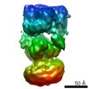
| 手法 | 電子顕微鏡法 / 単粒子再構成法 / クライオ電子顕微鏡法 / 解像度: 8.25 Å | ||||||
 データ登録者 データ登録者 | Herguedas, B. / Garcia-Nafria, J. / Fernandez-Leiro, R. / Greger, I.H. | ||||||
 引用 引用 | ジャーナル: Science / 年: 2016 タイトル: Structure and organization of heteromeric AMPA-type glutamate receptors. 著者: Beatriz Herguedas / Javier García-Nafría / Ondrej Cais / Rafael Fernández-Leiro / James Krieger / Hinze Ho / Ingo H Greger /  要旨: AMPA-type glutamate receptors (AMPARs), which are central mediators of rapid neurotransmission and synaptic plasticity, predominantly exist as heteromers of the subunits GluA1 to GluA4. Here we ...AMPA-type glutamate receptors (AMPARs), which are central mediators of rapid neurotransmission and synaptic plasticity, predominantly exist as heteromers of the subunits GluA1 to GluA4. Here we report the first AMPAR heteromer structures, which deviate substantially from existing GluA2 homomer structures. Crystal structures of the GluA2/3 and GluA2/4 N-terminal domains reveal a novel compact conformation with an alternating arrangement of the four subunits around a central axis. This organization is confirmed by cysteine cross-linking in full-length receptors, and it permitted us to determine the structure of an intact GluA2/3 receptor by cryogenic electron microscopy. Two models in the ligand-free state, at resolutions of 8.25 and 10.3 angstroms, exhibit substantial vertical compression and close associations between domain layers, reminiscent of N-methyl-D-aspartate receptors. Model 1 resembles a resting state and model 2 a desensitized state, thus providing snapshots of gating transitions in the nominal absence of ligand. Our data reveal organizational features of heteromeric AMPARs and provide a framework to decipher AMPAR architecture and signaling. | ||||||
| 履歴 |
|
- 構造の表示
構造の表示
| ムービー |
ムービービューア |
|---|---|
| 構造ビューア | 分子: MolmilJmol/JSmol |
- ダウンロードとリンク
-ダウンロード
| PDBx/mmCIF形式 | 5ide.cif.gz | 414.2 KB | 表示 | PDBx/mmCIF形式 |
|---|---|---|---|---|
| PDB形式 | pdb5ide.ent.gz | 254.4 KB | 表示 | PDB形式 |
| PDBx/mmJSON形式 | 5ide.json.gz | ツリー表示 | PDBx/mmJSON形式 | |
| その他 |  その他のダウンロード その他のダウンロード |
-検証レポート
| アーカイブディレクトリ | https://data.pdbj.org/pub/pdb/validation_reports/id/5ideftp://data.pdbj.org/pub/pdb/validation_reports/id/5ide | HTTPS FTP |
|---|
-関連構造データ


-リンク
 PDBj
PDBj


- 集合体
集合体
| 登録構造単位 | 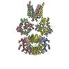
|
|---|---|
| 1 |
|
-要素
| #1: タンパク質 | GRIA2 / GluR-2 / AMPA-selective glutamate receptor 2 / GluR-B / GluR-K2 / Glutamate receptor ionotropic / ...GluR-2 / AMPA-selective glutamate receptor 2 / GluR-B / GluR-K2 / Glutamate receptor ionotropic / AMPA 2 / GluA2 分子量: 97663.188 Da / 分子数: 2 / 変異: N292C / 由来タイプ: 組換発現 詳細: The sequence corresponds to mature rat GluA2 (residues 22-883, isoform Flip, edited at R/G and Q/R sites) with a Myc tag after the first residue and the N292C mutation 由来: (組換発現) Rattus norvegicus (ドブネズミ) / 遺伝子: Gria2, Glur2 / プラスミド: pires2-EGFP / 細胞株 (発現宿主): HEK293 gntI- / 発現宿主:  Homo sapiens (ヒト) / 参照: UniProt: P19491 Homo sapiens (ヒト) / 参照: UniProt: P19491#2: タンパク質 | グルタミン酸受容体 / GluR-3 / AMPA-selective glutamate receptor 3 / GluR-C / GluR-K3 / Glutamate receptor ionotropic / ...GluR-3 / AMPA-selective glutamate receptor 3 / GluR-C / GluR-K3 / Glutamate receptor ionotropic / AMPA 3 / GluA3分子量: 99075.664 Da / 分子数: 2 / 変異: R439G, R265C / 由来タイプ: 組換発現 詳細: The sequence corresponds to the mature rat GluA3 subunit (residues 23-888, Flip isoform) with a Flag tag after the first residue and mutated at R439G and R265C 由来: (組換発現) Rattus norvegicus (ドブネズミ) / 遺伝子: Gria3, Glur3 / Variant: Flip / プラスミド: prk5 / 細胞株 (発現宿主): HEK293 gntI- / 発現宿主: Homo sapiens (ヒト) / 参照: UniProt: P19492 |
|---|
-実験情報
-実験
| 実験 | 手法: 電子顕微鏡法 |
|---|---|
| EM実験 | 試料の集合状態: PARTICLE / 3次元再構成法: 単粒子再構成法 |
- 試料調製
試料調製
| 構成要素 | 名称: AMPA GluA2/3 heterotetramer / タイプ: COMPLEX 詳細: Full-length GluA2/3 heterotetramer containing the A2_N292C and A3_265C mutations Entity ID: all / 由来: RECOMBINANT |
|---|---|
| 分子量 | 値: 0.4 MDa / 実験値: NO |
| 由来(天然) | 生物種: Rattus norvegicus (ドブネズミ) |
| 由来(組換発現) | 生物種: Homo sapiens (ヒト) / 細胞: HEK293 GntI- / プラスミド: prk5 and pIRES2-EGFP |
| 緩衝液 | pH: 7.4 / 詳細: 25 mM Tris pH 7.4, 0.25 % DDM, 150 mM NaCl |
| 試料 | 濃度: 0.03 mg/ml / 包埋: NO / シャドウイング: NO / 染色: NO / 凍結: YES |
| 試料支持 | グリッドの材料: COPPER / グリッドのサイズ: 300 divisions/in. / グリッドのタイプ: Quantifoil R1.2/1.3 |
| 急速凍結 | 装置: FEI VITROBOT MARK IV / 凍結剤: ETHANE / 湿度: 100 % / 凍結前の試料温度: 277 K / 詳細: Incubated for 1 minute, blotted for 3 seconds |
- 電子顕微鏡撮影
| 実験機器 |  モデル: Titan Krios / 画像提供: FEI Company |
|---|---|
| 顕微鏡 | モデル: FEI TITAN KRIOS |
| 電子銃 | 電子線源: FIELD EMISSION GUN / 加速電圧: 300 kV / 照射モード: OTHER |
| 電子レンズ | モード: BRIGHT FIELDBright-field microscopy / 倍率(補正後): 28409 X / 最大 デフォーカス(公称値): 4000 nm / 最小 デフォーカス(公称値): 1000 nm / Cs: 2.7 mm / C2レンズ絞り径: 70 µm |
| 試料ホルダ | 凍結剤: NITROGEN 試料ホルダーモデル: FEI TITAN KRIOS AUTOGRID HOLDER |
| 撮影 | 平均露光時間: 25 sec. / 電子線照射量: 40 e/Å2 / 検出モード: COUNTING フィルム・検出器のモデル: GATAN K2 SUMMIT (4k x 4k) 撮影したグリッド数: 2 / 実像数: 980 |
| 電子光学装置 | エネルギーフィルター名称: GIF Quantum / エネルギーフィルター 上限: 20 eV / エネルギーフィルター 下限: 0 eV |
| 画像スキャン | 動画フレーム数/画像: 20 / 利用したフレーム数/画像: 1-20 |
- 解析
解析
| EMソフトウェア |
| |||||||||||||||||||||||||||||||||||||||||||||
|---|---|---|---|---|---|---|---|---|---|---|---|---|---|---|---|---|---|---|---|---|---|---|---|---|---|---|---|---|---|---|---|---|---|---|---|---|---|---|---|---|---|---|---|---|---|---|
| CTF補正 | タイプ: PHASE FLIPPING AND AMPLITUDE CORRECTION | |||||||||||||||||||||||||||||||||||||||||||||
| 粒子像の選択 | 選択した粒子像数: 107939 | |||||||||||||||||||||||||||||||||||||||||||||
| 対称性 | 点対称性: C1 (非対称) | |||||||||||||||||||||||||||||||||||||||||||||
| 3次元再構成 | 解像度: 8.25 Å / 解像度の算出法: FSC 0.143 CUT-OFF / 粒子像の数: 25238 / 対称性のタイプ: POINT | |||||||||||||||||||||||||||||||||||||||||||||
| 原子モデル構築 | プロトコル: RIGID BODY FIT / 空間: REAL 詳細: For GluA2 chains (A,C), 2 copies of GluA2NTD (3HSY) and two copies of GluA2 LBD (1FTO) were fitted. For GluA3 chains (B,D), 2 copies of GluA3NTD (3O21) and two copies of GluA2 LBD (3UA8) were ...詳細: For GluA2 chains (A,C), 2 copies of GluA2NTD (3HSY) and two copies of GluA2 LBD (1FTO) were fitted. For GluA3 chains (B,D), 2 copies of GluA3NTD (3O21) and two copies of GluA2 LBD (3UA8) were fitted.For the TMD region, the 4 chains of 3KG2 TMD(residues 509-544 594-629 784-817) were fitted as a rigid body. After fitting the sequence was corrected including mutations and side chains were removed. | |||||||||||||||||||||||||||||||||||||||||||||
| 原子モデル構築 | 3D fitting-ID: 1 / Source name: PDB / タイプ: experimental model
|