 ムービー
ムービー コントローラー


+ データを開く

- 基本情報
基本情報
| 登録情報 | データベース: EMDB / ID: EMD-5280 | |||||||||
|---|---|---|---|---|---|---|---|---|---|---|
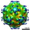
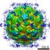
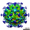
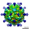
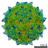
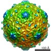
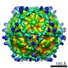
| タイトル | Poliovirus 135S particle and P1 Fab complex at 12-angs. resolution | |||||||||
 マップデータ マップデータ | This is a map of poliovirus 135S and P1 Fab complex at 12-ang resolution | |||||||||
 試料 試料 |
| |||||||||
 キーワード キーワード | picornavirus / viral cell entry / viral uncoating / virus-antibody complex / virus-Fab complex / virus disassembly / virus uncoating / virus conformational transitions / monospecific antibody | |||||||||
| 生物種 |   Human poliovirus 1 Mahoney (ポリオウイルス) Human poliovirus 1 Mahoney (ポリオウイルス) | |||||||||
| 手法 | 単粒子再構成法 / クライオ電子顕微鏡法 / 解像度: 12.0 Å | |||||||||
 データ登録者 データ登録者 | Lin J / Cheng N / Chow M / Filman DJ / Steven AC / Hogle JM / Belnap DM | |||||||||
 引用 引用 | ジャーナル: J Virol / 年: 2011 タイトル: An externalized polypeptide partitions between two distinct sites on genome-released poliovirus particles. 著者: Jun Lin / Naiqian Cheng / Marie Chow / David J Filman / Alasdair C Steven / James M Hogle / David M Belnap /  要旨: During cell entry, native poliovirus (160S) converts to a cell-entry intermediate (135S) particle, resulting in the externalization of capsid proteins VP4 and the amino terminus of VP1 (residues 1 to ...During cell entry, native poliovirus (160S) converts to a cell-entry intermediate (135S) particle, resulting in the externalization of capsid proteins VP4 and the amino terminus of VP1 (residues 1 to 53). Externalization of these entities is followed by release of the RNA genome (uncoating), leaving an empty (80S) particle. The antigen-binding fragment (Fab) of a monospecific peptide 1 (P1) antibody, which was raised against a peptide corresponding to amino-terminal residues 24 to 40 of VP1, was utilized to track the location of the amino terminus of VP1 in the 135S and 80S states of poliovirus particles via cryogenic electron microscopy (cryo-EM) and three-dimensional image reconstruction. On 135S, P1 Fabs bind to a prominent feature on the external surface known as the "propeller tip." In contrast, our initial 80S-P1 reconstruction showed P1 Fabs also binding to a second site, at least 50 Å distant, at the icosahedral 2-fold axes. Further analysis showed that the overall population of 80S-P1 particles consisted of three kinds of capsids: those with P1 Fabs bound only at the propeller tips, P1 Fabs bound only at the 2-fold axes, or P1 Fabs simultaneously bound at both positions. Our results indicate that, in 80S particles, a significant fraction of VP1 can deviate from icosahedral symmetry. Hence, this portion of VP1 does not change conformation synchronously when switching from the 135S state. These conclusions are compatible with previous observations of multiple conformations of the 80S state and suggest that movement of the amino terminus of VP1 has a role in uncoating. Similar deviations from icosahedral symmetry may be biologically significant during other viral transitions. | |||||||||
| 履歴 |
|
- 構造の表示
構造の表示
| ムービー |
 ムービービューア ムービービューア |
|---|---|
| 構造ビューア | EMマップ: SurfViewMolmilJmol/JSmol |
| 添付画像 |

- ダウンロードとリンク
-EMDBアーカイブ
| マップデータ | emd_5280.map.gz | 34.7 MB | EMDBマップデータ形式 | |
|---|---|---|---|---|
| ヘッダ (付随情報) | emd-5280-v30.xmlemd-5280.xml | 10 KB 10 KB | 表示 表示 | EMDBヘッダ |
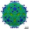
| 画像 | 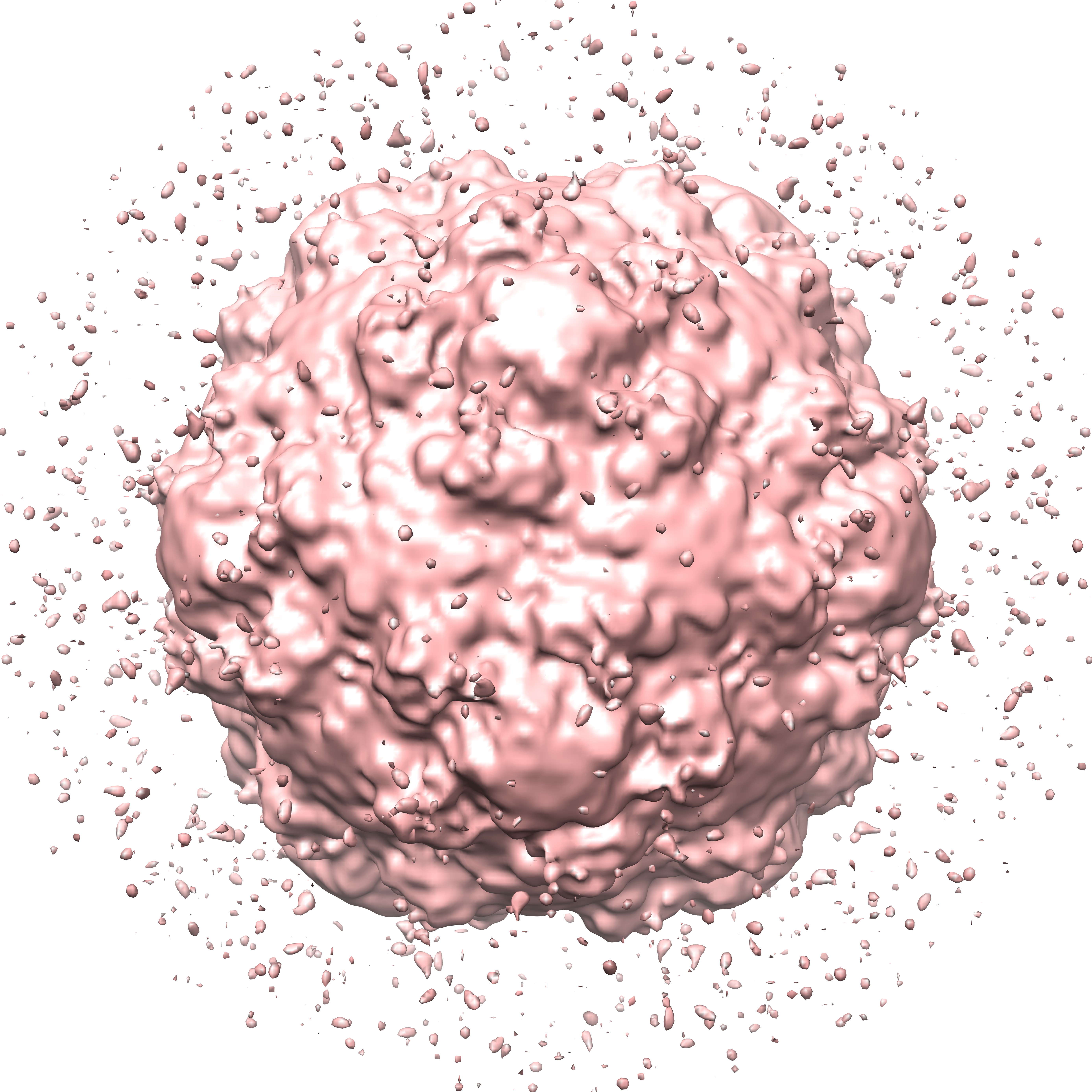 emd_5280_1.jpg emd_5280_1.jpg | 2.1 MB | ||
| アーカイブディレクトリ |  http://ftp.pdbj.org/pub/emdb/structures/EMD-5280ftp://ftp.pdbj.org/pub/emdb/structures/EMD-5280 http://ftp.pdbj.org/pub/emdb/structures/EMD-5280ftp://ftp.pdbj.org/pub/emdb/structures/EMD-5280 | HTTPS FTP |
-検証レポート
| 文書・要旨 | emd_5280_validation.pdf.gz | 78.2 KB | 表示 | EMDB検証レポート |
|---|---|---|---|---|
| 文書・詳細版 | emd_5280_full_validation.pdf.gz | 77.3 KB | 表示 | |
| XML形式データ | emd_5280_validation.xml.gz | 493 B | 表示 | |
| アーカイブディレクトリ | https://ftp.pdbj.org/pub/emdb/validation_reports/EMD-5280ftp://ftp.pdbj.org/pub/emdb/validation_reports/EMD-5280 | HTTPS FTP |
-関連構造データ

-リンク
| EMDBのページ | EMDB (EBI/PDBe) / EMDataResource |
|---|---|
| 「今月の分子」の関連する項目 |

-マップ
| ファイル | ダウンロード / ファイル: emd_5280.map.gz / 形式: CCP4 / 大きさ: 70.9 MB / タイプ: IMAGE STORED AS FLOATING POINT NUMBER (4 BYTES) | ||||||||||||||||||||||||||||||||||||||||||||||||||||||||||||||||||||
|---|---|---|---|---|---|---|---|---|---|---|---|---|---|---|---|---|---|---|---|---|---|---|---|---|---|---|---|---|---|---|---|---|---|---|---|---|---|---|---|---|---|---|---|---|---|---|---|---|---|---|---|---|---|---|---|---|---|---|---|---|---|---|---|---|---|---|---|---|---|
| 注釈 | This is a map of poliovirus 135S and P1 Fab complex at 12-ang resolution | ||||||||||||||||||||||||||||||||||||||||||||||||||||||||||||||||||||
| ボクセルのサイズ | X=Y=Z: 1.83 Å | ||||||||||||||||||||||||||||||||||||||||||||||||||||||||||||||||||||
| 密度 |
| ||||||||||||||||||||||||||||||||||||||||||||||||||||||||||||||||||||
| 対称性 | 空間群: 1 | ||||||||||||||||||||||||||||||||||||||||||||||||||||||||||||||||||||
| 詳細 | EMDB XML:
CCP4マップ ヘッダ情報:
| ||||||||||||||||||||||||||||||||||||||||||||||||||||||||||||||||||||
-添付データ
- 試料の構成要素
-全体 : Poliovirus 135S particle and P1(monospecific antibody) Fab complex
| 全体 | 名称: Poliovirus 135S particle and P1(monospecific antibody) Fab complex |
|---|---|
| 要素 |
|
-超分子 #1000: Poliovirus 135S particle and P1(monospecific antibody) Fab complex
| 超分子 | 名称: Poliovirus 135S particle and P1(monospecific antibody) Fab complex タイプ: sample / ID: 1000 / 集合状態: 135S particle icosahedral with Fab / Number unique components: 2 |
|---|
-超分子 #1: Human poliovirus 1 Mahoney
| 超分子 | 名称: Human poliovirus 1 Mahoney / タイプ: virus / ID: 1 / Name.synonym: poliovirus 135S 詳細: native virus 160S is converted by heat-treatment to 135S NCBI-ID: 12081 / 生物種: Human poliovirus 1 Mahoney / データベース: NCBI / ウイルスタイプ: VIRION / ウイルス・単離状態: STRAIN / ウイルス・エンベロープ: No / ウイルス・中空状態: No / Syn species name: poliovirus 135S |
|---|---|
| 宿主 | 生物種:  Homo sapiens (ヒト) / 別称: VERTEBRATES Homo sapiens (ヒト) / 別称: VERTEBRATES |
| ウイルス殻 | Shell ID: 1 / 直径: 340 Å / T番号(三角分割数): 1 |
-実験情報
-構造解析
| 手法 | クライオ電子顕微鏡法 |
|---|---|
 解析 解析 | 単粒子再構成法 |
| 試料の集合状態 | particle |
-試料調製
| 緩衝液 | pH: 7.5 / 詳細: 20 mM Tris, 2 mM CaCl2 |
|---|---|
| 凍結 | 凍結剤: ETHANE / 装置: OTHER 詳細: Vitrification carried out in ambient atmosphere. Ethane cooled by liquid nitrogen. 手法: Blotted manually before plunging |
- 電子顕微鏡法
| 顕微鏡 | FEI/PHILIPS CM200FEG |
|---|---|
| アライメント法 | Legacy - 非点収差: Bsoft |
| 日付 | 1999年7月15日 |
| 撮影 | カテゴリ: FILM / フィルム・検出器のモデル: KODAK SO-163 FILM / デジタル化 - スキャナー: ZEISS SCAI / デジタル化 - サンプリング間隔: 7 µm / 実像数: 14 / 平均電子線量: 10 e/Å2 / 詳細: Defocal pairs were used. / ビット/ピクセル: 8 |
| Tilt angle min | 0 |
| Tilt angle max | 0 |
| 電子線 | 加速電圧: 120 kV / 電子線源:  FIELD EMISSION GUN FIELD EMISSION GUN |
| 電子光学系 | 倍率(補正後): 37752 / 照射モード: FLOOD BEAM / 撮影モード: BRIGHT FIELD / Cs: 2 mm / 最大 デフォーカス(公称値): 2.24 µm / 最小 デフォーカス(公称値): 0.81 µm / 倍率(公称値): 38000 |
| 試料ステージ | 試料ホルダー: Side entry liquid nitrogen-cooled cryo specimen holder 試料ホルダーモデル: GATAN LIQUID NITROGEN |
-画像解析
| CTF補正 | 詳細: CTF and decay correction of each particle |
|---|---|
| 最終 再構成 | アルゴリズム: OTHER / 解像度のタイプ: BY AUTHOR / 解像度: 12.0 Å / 解像度の算出法: FSC 0.5 CUT-OFF / ソフトウェア - 名称: EM3DR2 詳細: Reconstruction computed from focal pairs. Pairs not summed for reconstruction calculation. 使用した粒子像数: 10160 |
