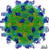
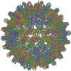
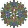
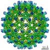
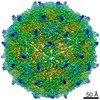
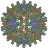
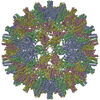
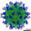
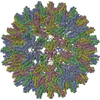
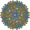
ジャーナル: PLoS One / 年: 2013 タイトル: 3.5Å cryoEM structure of hepatitis B virus core assembled from full-length core protein. 著者: Xuekui Yu / Lei Jin / Jonathan Jih / Chiaho Shih / Z Hong Zhou / 要旨: The capsid shell of infectious hepatitis B virus (HBV) is composed of 240 copies of a single protein called HBV core antigen (HBc). An atomic model of a core assembled from truncated HBc was ...The capsid shell of infectious hepatitis B virus (HBV) is composed of 240 copies of a single protein called HBV core antigen (HBc). An atomic model of a core assembled from truncated HBc was determined previously by X-ray crystallography. In an attempt to obtain atomic structural information of HBV core in a near native, non-crystalline environment, we reconstructed a 3.5Å-resolution structure of a recombinant core assembled from full-length HBc by cryo electron microscopy (cryoEM) and derived an atomic model. The structure shows that the 240 molecules of full-length HBc form a core with two layers. The outer layer, composed of the N-terminal assembly domain, is similar to the crystal structure of the truncated HBc, but has three differences. First, unlike the crystal structure, our cryoEM structure shows no disulfide bond between the Cys61 residues of the two subunits within the dimer building block, indicating such bond is not required for core formation. Second, our cryoEM structure reveals up to four more residues in the linker region (amino acids 140-149). Third, the loops in the cryoEM structures containing this linker region in subunits B and C are oriented differently (~30° and ~90°) from their counterparts in the crystal structure. The inner layer, composed of the C-terminal arginine-rich domain (ARD) and the ARD-bound RNAs, is partially-ordered and connected with the outer layer through linkers positioned around the two-fold axes. Weak densities emanate from the rims of positively charged channels through the icosahedral three-fold and local three-fold axes. We attribute these densities to the exposed portions of some ARDs, thus explaining ARD's accessibility by proteases and antibodies. Our data supports a role of ARD in mediating communication between inside and outside of the core during HBV maturation and envelopment.
 ムービー
ムービー コントローラー


 データを開く
 基本情報
基本情報 要素
要素 キーワード
キーワード VIRUS (ウイルス) / Hepatitis B virus core antigen (HBc) / no Cys61 intermolecular disulfide bond
 機能・相同性情報

 データ登録者
 引用
引用
 構造の表示
構造の表示 ダウンロードとリンク
 その他のダウンロード
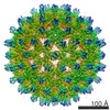

 PDBj
PDBj

 集合体
集合体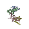


 試料調製
試料調製 電子顕微鏡撮影
 解析
解析