ジャーナル: Sci Rep / 年: 2017 タイトル: Visualizing Adsorption of Cyanophage P-SSP7 onto Marine Prochlorococcus. 著者: Kazuyoshi Murata / Qinfen Zhang / Jesús Gerardo Galaz-Montoya / Caroline Fu / Maureen L Coleman / Marcia S Osburne / Michael F Schmid / Matthew B Sullivan / Sallie W Chisholm / Wah Chiu / 要旨: Marine cyanobacteria perform roughly a quarter of global carbon fixation, and cyanophages that infect them liberate some of this carbon during infection and cell lysis. Studies of the cyanobacterium ...Marine cyanobacteria perform roughly a quarter of global carbon fixation, and cyanophages that infect them liberate some of this carbon during infection and cell lysis. Studies of the cyanobacterium Prochlorococcus MED4 and its associated cyanophage P-SSP7 have revealed complex gene expression dynamics once infection has begun, but the initial cyanophage-host interactions remain poorly understood. Here, we used single particle cryo-electron tomography (cryo-ET) to investigate cyanophage-host interactions in this model system, based on 170 cyanophage-to-host adsorption events. Subtomogram classification and averaging revealed three main conformations characterized by different angles between the phage tail and the cell surface. Namely, phage tails were (i) parallel to, (ii) ~45 degrees to, or (iii) perpendicular to the cell surface. Furthermore, different conformations of phage tail fibers correlated with the aforementioned orientations of the tails. We also observed density beyond the tail tip in vertically-oriented phages that had penetrated the cell wall, capturing the final stage of adsorption. Together, our data provide a quantitative characterization of the orientation of phages as they adsorb onto cells, and suggest that cyanophages that abut their cellular targets are only transiently in the "perpendicular" orientation required for successful infection.
 ムービー
ムービー コントローラー


 データを開く
 基本情報
基本情報 マップデータ
 試料
試料 キーワード
キーワード cryo-electron tomography /
  Prochlorococcus phage P-SSP7 (ファージ)
 データ登録者
 引用
引用


 構造の表示
構造の表示 ムービービューア
 ダウンロードとリンク
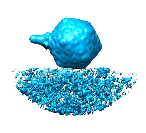 EMD-3131.jpg
 http://ftp.pdbj.org/pub/emdb/structures/EMD-3131

 試料の構成要素
 解析
解析 電子顕微鏡法