 ムービー
ムービー コントローラー


+ データを開く

- 基本情報
基本情報
| 登録情報 | データベース: PDB / ID: 1ry1 | ||||||
|---|---|---|---|---|---|---|---|
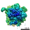
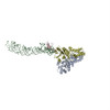
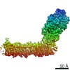
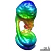
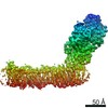
| タイトル | Structure of the signal recognition particle interacting with the elongation-arrested ribosome | ||||||
 要素 要素 |
| ||||||
 キーワード キーワード |  TRANSLATION (翻訳 (生物学)) / signal recognition particle (シグナル認識粒子) / RNA binding (リボ核酸) TRANSLATION (翻訳 (生物学)) / signal recognition particle (シグナル認識粒子) / RNA binding (リボ核酸) | ||||||
| 機能・相同性 |  機能・相同性情報signal recognition particle receptor complex / SRP-dependent cotranslational protein targeting to membrane / SRP-dependent cotranslational protein targeting to membrane, signal sequence recognition / endoplasmic reticulum signal peptide binding / signal recognition particle, endoplasmic reticulum targeting / signal recognition particle binding / granulocyte differentiation / absorption of visible light / negative regulation of translational elongation / G protein-coupled photoreceptor activity ...signal recognition particle receptor complex / SRP-dependent cotranslational protein targeting to membrane / SRP-dependent cotranslational protein targeting to membrane, signal sequence recognition / endoplasmic reticulum signal peptide binding / signal recognition particle, endoplasmic reticulum targeting / signal recognition particle binding / granulocyte differentiation / absorption of visible light / negative regulation of translational elongation / G protein-coupled photoreceptor activity / シグナル認識粒子 / photoreceptor inner segment membrane / rhodopsin mediated signaling pathway / 11-cis retinal binding / cotranslational protein targeting to membrane / signal-recognition-particle GTPase / protein targeting to ER / SRP-dependent cotranslational protein targeting to membrane, translocation / 7S RNA binding / exocrine pancreas development / SRP-dependent cotranslational protein targeting to membrane / photoreceptor outer segment membrane / SRP-dependent cotranslational protein targeting to membrane / ribonucleoprotein complex binding / 視覚 / 好中球 / photoreceptor disc membrane / GDP binding / secretory granule lumen / ficolin-1-rich granule lumen / nuclear body / nuclear speck / GTPase activity / Neutrophil degranulation / GTP binding / 核小体 / 小胞体 / ATP hydrolysis activity / RNA binding / extracellular region / 生体膜 / metal ion binding / 細胞核 / 細胞膜 / 細胞質基質 / 細胞質 機能・相同性情報signal recognition particle receptor complex / SRP-dependent cotranslational protein targeting to membrane / SRP-dependent cotranslational protein targeting to membrane, signal sequence recognition / endoplasmic reticulum signal peptide binding / signal recognition particle, endoplasmic reticulum targeting / signal recognition particle binding / granulocyte differentiation / absorption of visible light / negative regulation of translational elongation / G protein-coupled photoreceptor activity ...signal recognition particle receptor complex / SRP-dependent cotranslational protein targeting to membrane / SRP-dependent cotranslational protein targeting to membrane, signal sequence recognition / endoplasmic reticulum signal peptide binding / signal recognition particle, endoplasmic reticulum targeting / signal recognition particle binding / granulocyte differentiation / absorption of visible light / negative regulation of translational elongation / G protein-coupled photoreceptor activity / シグナル認識粒子 / photoreceptor inner segment membrane / rhodopsin mediated signaling pathway / 11-cis retinal binding / cotranslational protein targeting to membrane / signal-recognition-particle GTPase / protein targeting to ER / SRP-dependent cotranslational protein targeting to membrane, translocation / 7S RNA binding / exocrine pancreas development / SRP-dependent cotranslational protein targeting to membrane / photoreceptor outer segment membrane / SRP-dependent cotranslational protein targeting to membrane / ribonucleoprotein complex binding / 視覚 / 好中球 / photoreceptor disc membrane / GDP binding / secretory granule lumen / ficolin-1-rich granule lumen / nuclear body / nuclear speck / GTPase activity / Neutrophil degranulation / GTP binding / 核小体 / 小胞体 / ATP hydrolysis activity / RNA binding / extracellular region / 生体膜 / metal ion binding / 細胞核 / 細胞膜 / 細胞質基質 / 細胞質類似検索 - 分子機能 | ||||||
| 生物種 |  Canis lupus familiaris (イヌ) Canis lupus familiaris (イヌ) Triticum aestivum (コムギ) Triticum aestivum (コムギ) | ||||||
| 手法 | 電子顕微鏡法 / 単粒子再構成法 / クライオ電子顕微鏡法 / 解像度: 12 Å | ||||||
 データ登録者 データ登録者 | Halic, M. / Becker, T. / Pool, M.R. / Spahn, C.M. / Grassucci, R.A. / Frank, J. / Beckmann, R. | ||||||
 引用 引用 | ジャーナル: Nature / 年: 2004 タイトル: Structure of the signal recognition particle interacting with the elongation-arrested ribosome. 著者: Mario Halic / Thomas Becker / Martin R Pool / Christian M T Spahn / Robert A Grassucci / Joachim Frank / Roland Beckmann /  要旨: Cotranslational translocation of proteins across or into membranes is a vital process in all kingdoms of life. It requires that the translating ribosome be targeted to the membrane by the signal ...Cotranslational translocation of proteins across or into membranes is a vital process in all kingdoms of life. It requires that the translating ribosome be targeted to the membrane by the signal recognition particle (SRP), an evolutionarily conserved ribonucleoprotein particle. SRP recognizes signal sequences of nascent protein chains emerging from the ribosome. Subsequent binding of SRP leads to a pause in peptide elongation and to the ribosome docking to the membrane-bound SRP receptor. Here we present the structure of a targeting complex consisting of mammalian SRP bound to an active 80S ribosome carrying a signal sequence. This structure, solved to 12 A by cryo-electron microscopy, enables us to generate a molecular model of SRP in its functional conformation. The model shows how the S domain of SRP contacts the large ribosomal subunit at the nascent chain exit site to bind the signal sequence, and that the Alu domain reaches into the elongation-factor-binding site of the ribosome, explaining its elongation arrest activity. | ||||||
| 履歴 |
|
- 構造の表示
構造の表示
| ムービー |
ムービービューア |
|---|---|
| 構造ビューア | 分子: MolmilJmol/JSmol |
- ダウンロードとリンク
-ダウンロード
| PDBx/mmCIF形式 | 1ry1.cif.gz | 310 KB | 表示 | PDBx/mmCIF形式 |
|---|---|---|---|---|
| PDB形式 | pdb1ry1.ent.gz | 231.9 KB | 表示 | PDB形式 |
| PDBx/mmJSON形式 | 1ry1.json.gz | ツリー表示 | PDBx/mmJSON形式 | |
| その他 |  その他のダウンロード その他のダウンロード |
-検証レポート
| アーカイブディレクトリ | https://data.pdbj.org/pub/pdb/validation_reports/ry/1ry1ftp://data.pdbj.org/pub/pdb/validation_reports/ry/1ry1 | HTTPS FTP |
|---|
-関連構造データ
-リンク
 PDBj
PDBj


- 集合体
集合体
| 登録構造単位 | 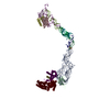
|
|---|---|
| 1 |
|
-要素
-RNA鎖 , 8種, 8分子 EAMNOPQR
| #1: RNA鎖 | 分子量: 16277.622 Da / 分子数: 1 / 由来タイプ: 天然 / 由来: (天然) Canis lupus familiaris (イヌ) |
|---|---|
| #2: RNA鎖 | 分子量: 41566.715 Da / 分子数: 1 / 由来タイプ: 天然 / 由来: (天然) Canis lupus familiaris (イヌ) |
| #3: RNA鎖 | Signal recognition particle RNA 分子量: 8784.278 Da / 分子数: 1 / 由来タイプ: 天然 / 由来: (天然) Canis lupus familiaris (イヌ) |
| #4: RNA鎖 | Signal recognition particle RNA 分子量: 9807.837 Da / 分子数: 1 / 由来タイプ: 天然 / 由来: (天然) Canis lupus familiaris (イヌ) |
| #5: RNA鎖 | Signal recognition particle RNA 分子量: 7695.542 Da / 分子数: 1 / 由来タイプ: 天然 / 由来: (天然) Canis lupus familiaris (イヌ) |
| #6: RNA鎖 | Signal recognition particle RNA 分子量: 6325.847 Da / 分子数: 1 / 由来タイプ: 天然 / 由来: (天然) Canis lupus familiaris (イヌ) |
| #7: RNA鎖 | Signal recognition particle RNA 分子量: 3844.320 Da / 分子数: 1 / 由来タイプ: 天然 / 由来: (天然) Canis lupus familiaris (イヌ) |
| #8: RNA鎖 | Signal recognition particle RNA 分子量: 3804.296 Da / 分子数: 1 / 由来タイプ: 天然 / 由来: (天然) Canis lupus familiaris (イヌ) |
-タンパク質 , 5種, 5分子 CDBUW
| #9: タンパク質 | Signal recognition particle 9 分子量: 9996.567 Da / 分子数: 1 / 由来タイプ: 天然 / 由来: (天然) Canis lupus familiaris (イヌ) / 属: Homoヒト属 / 参照: UniProt: P49458*PLUS |
|---|---|
| #10: タンパク質 | 分子量: 12114.235 Da / 分子数: 1 / 由来タイプ: 天然 / 由来: (天然) Canis lupus familiaris (イヌ) / 属: Homoヒト属 / 参照: UniProt: P37108 |
| #11: タンパク質 | 分子量: 12561.688 Da / 分子数: 1 / 由来タイプ: 天然 / 由来: (天然) Canis lupus familiaris (イヌ) / 属: Homoヒト属 / 参照: UniProt: P09132*PLUS |
| #12: タンパク質 | 分子量: 32472.508 Da / 分子数: 1 / 由来タイプ: 天然 / 由来: (天然) Canis lupus familiaris (イヌ) / 属: Thermusサーマス属 / 参照: UniProt: O07347*PLUS |
| #13: タンパク質 | 分子量: 12473.440 Da / 分子数: 1 / 由来タイプ: 天然 / 由来: (天然) Canis lupus familiaris (イヌ) / 属: Mus / 参照: UniProt: P14576*PLUS |
-タンパク質・ペプチド , 1種, 1分子 S
| #14: タンパク質・ペプチド | 分子量: 2121.542 Da / 分子数: 1 / 由来タイプ: 合成 / 由来: (合成) Triticum aestivum (コムギ) / 参照: UniProt: O62798*PLUS |
|---|
-詳細
| 配列の詳細 | This structure has been modeled using polymer sequences from other organisms, including Homo ...This structure has been modeled using polymer sequences from other organisms, including Homo sapiens (chains C,D,E), Thermus aquaticus (chain U), Mus musculus (chain W), and Tursiops truncatus (chain S). |
|---|
-実験情報
-実験
| 実験 | 手法: 電子顕微鏡法 |
|---|---|
| EM実験 | 試料の集合状態: PARTICLE / 3次元再構成法: 単粒子再構成法 |
- 試料調製
試料調製
| 構成要素 |
| ||||||||||||||||||||||||
|---|---|---|---|---|---|---|---|---|---|---|---|---|---|---|---|---|---|---|---|---|---|---|---|---|---|
| 由来(天然) |
| ||||||||||||||||||||||||
| 緩衝液 | pH: 7.4 | ||||||||||||||||||||||||
| 試料 | 包埋: NO / シャドウイング: NO / 染色: NO / 凍結: YES | ||||||||||||||||||||||||
| 試料支持 | 詳細: HOLEY CARBON | ||||||||||||||||||||||||
| 急速凍結 | 凍結剤: ETHANE / 詳細: PLUNGED INTO ETHANE |
- 電子顕微鏡撮影
| 実験機器 |  モデル: Tecnai F20 / 画像提供: FEI Company |
|---|---|
| 顕微鏡 | モデル: FEI TECNAI F20 / 日付: 2000年1月1日 詳細: SAMPLES WERE MAINTAINED AT LIQUID NITROGEN TEMPERATURES IN THE ELECTRON MICROSCOPE. |
| 電子銃 | 電子線源: FIELD EMISSION GUN / 加速電圧: 160 kV / 照射モード: FLOOD BEAM |
| 電子レンズ | モード: BRIGHT FIELDBright-field microscopy / 倍率(公称値): 51000 X / 倍率(補正後): 52000 X / 最大 デフォーカス(公称値): 45000 nm / 最小 デフォーカス(公称値): 10000 nm / Cs: 2 mm |
| 試料ホルダ | 温度: 95 K / 傾斜角・最大: 0 ° / 傾斜角・最小: 0 ° |
| 撮影 | 電子線照射量: 10 e/Å2 / フィルム・検出器のモデル: KODAK SO-163 FILM |
| 画像スキャン | デジタル画像の数: 75 |
- 解析
解析
| EMソフトウェア |
| ||||||||||||
|---|---|---|---|---|---|---|---|---|---|---|---|---|---|
| 対称性 | 点対称性: C1 (非対称) | ||||||||||||
| 3次元再構成 | 解像度: 12 Å / 解像度の算出法: FSC 0.5 CUT-OFF 詳細: The chains M, N, O, P, Q and R are fragments of a double helical strand of RNA. The author maintains that some of the residues could not be modeled correctly due to limited resolution in this region. 対称性のタイプ: POINT | ||||||||||||
| 精密化 | 最高解像度: 12 Å | ||||||||||||
| 精密化ステップ | サイクル: LAST / 最高解像度: 12 Å
|
